Academic Profile
Statistics
Similar Authors
Papers on arXiv
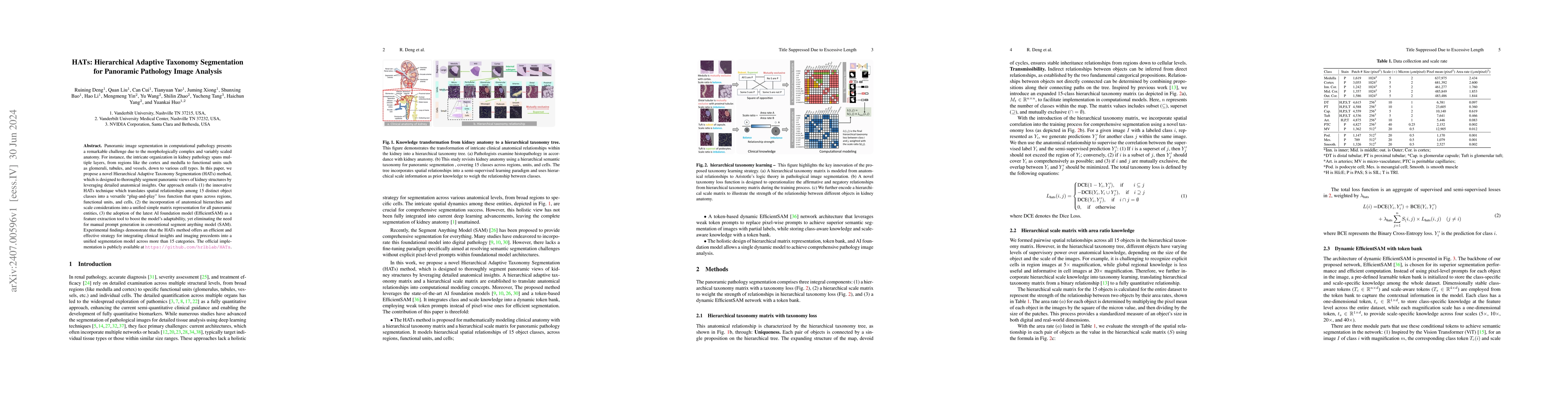
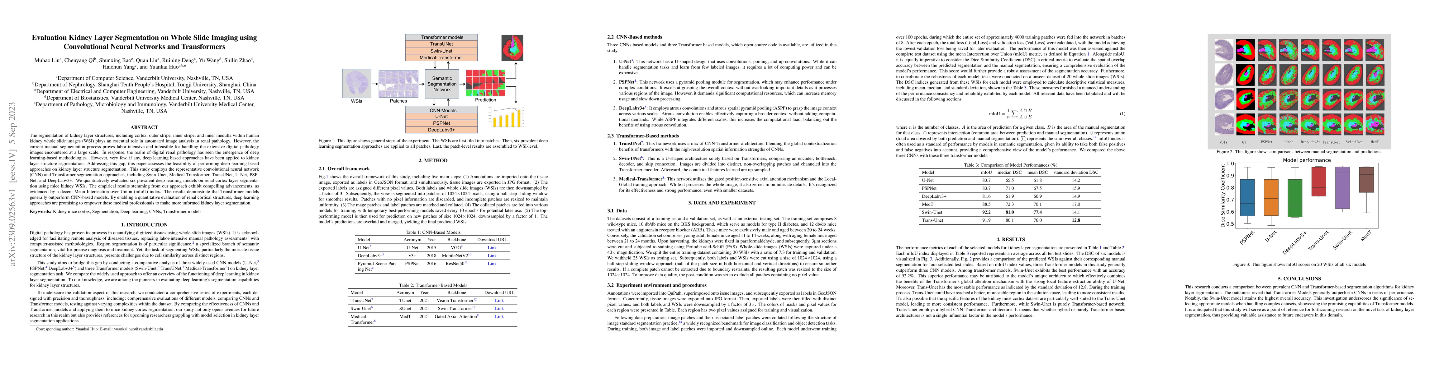
Panoramic image segmentation in computational pathology presents a remarkable challenge due to the morphologically complex and variably scaled anatomy. For instance, the intricate organization in kidn...
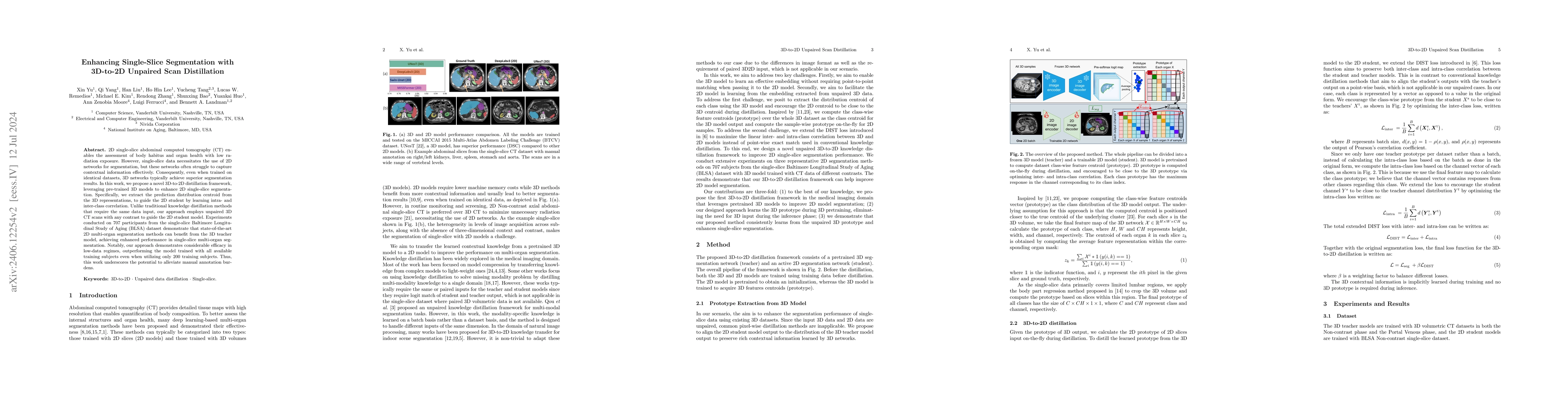
2D single-slice abdominal computed tomography (CT) enables the assessment of body habitus and organ health with low radiation exposure. However, single-slice data necessitates the use of 2D networks f...
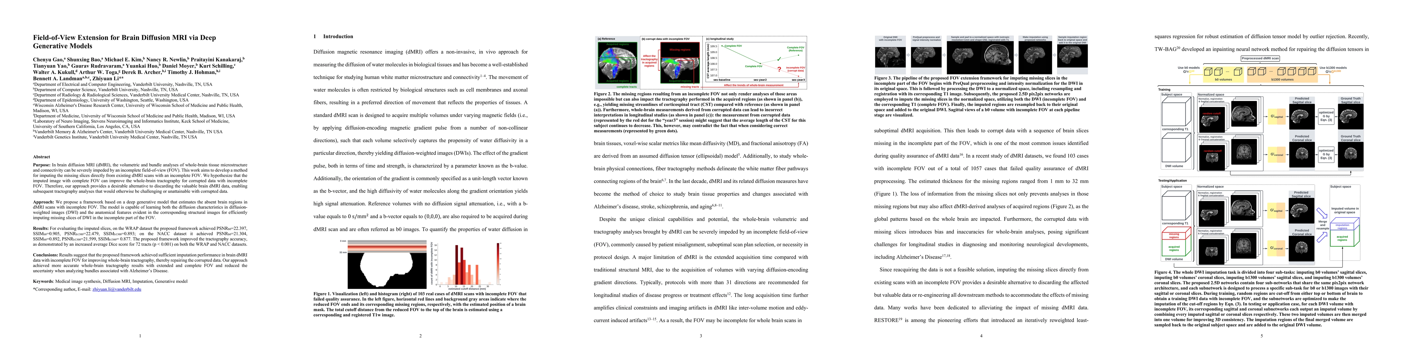
Purpose: In diffusion MRI (dMRI), the volumetric and bundle analyses of whole-brain tissue microstructure and connectivity can be severely impeded by an incomplete field-of-view (FOV). This work aim...
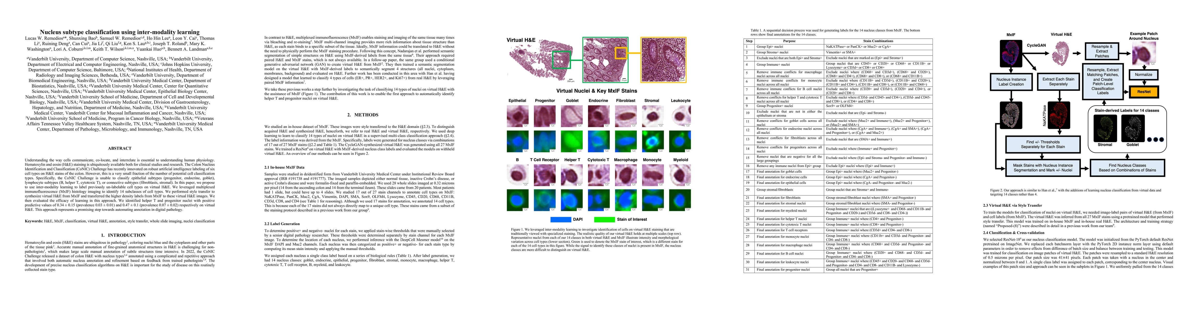
Understanding the way cells communicate, co-locate, and interrelate is essential to understanding human physiology. Hematoxylin and eosin (H&E) staining is ubiquitously available both for clinical s...
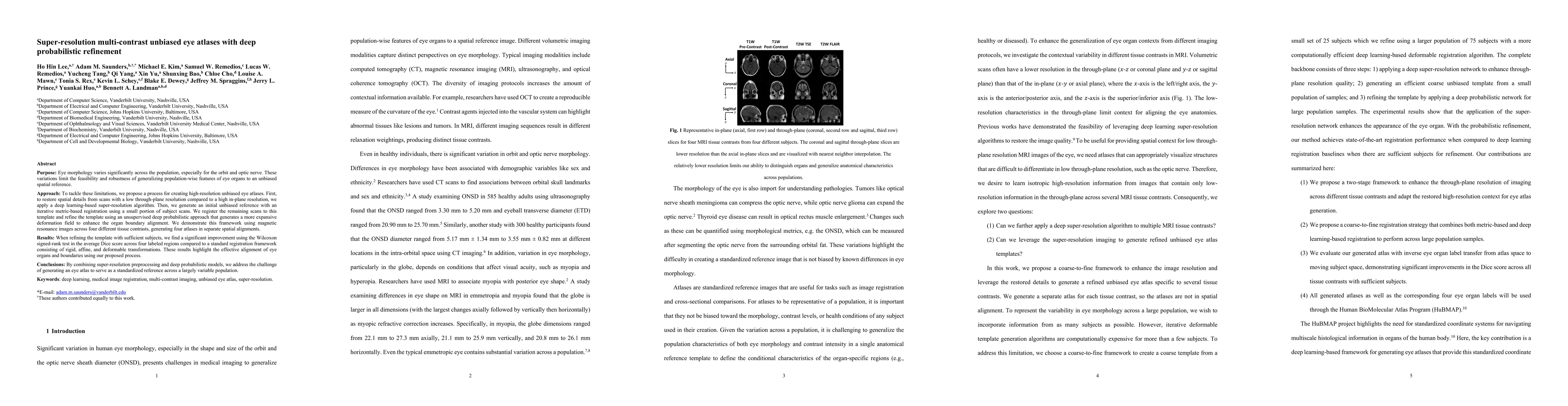
Purpose: Eye morphology varies significantly across the population, especially for the orbit and optic nerve. These variations limit the feasibility and robustness of generalizing population-wise fe...
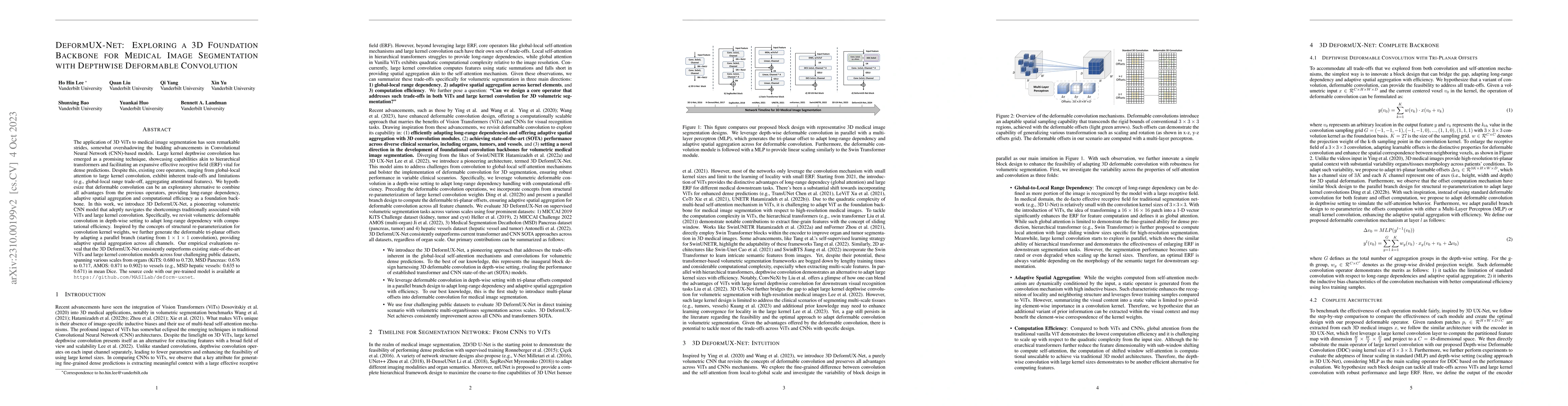
The application of 3D ViTs to medical image segmentation has seen remarkable strides, somewhat overshadowing the budding advancements in Convolutional Neural Network (CNN)-based models. Large kernel...
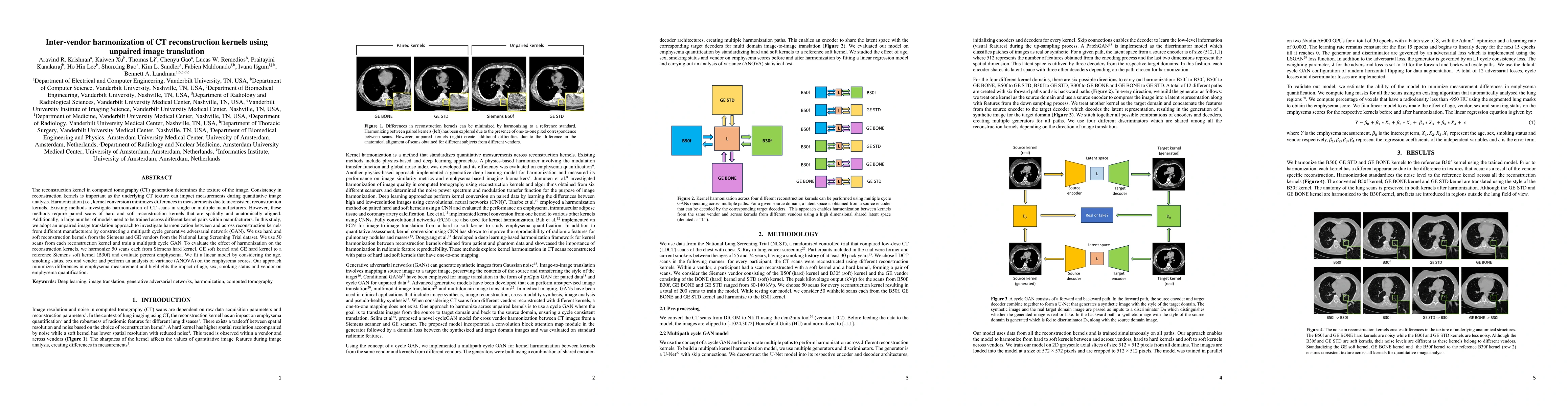
The reconstruction kernel in computed tomography (CT) generation determines the texture of the image. Consistency in reconstruction kernels is important as the underlying CT texture can impact measu...
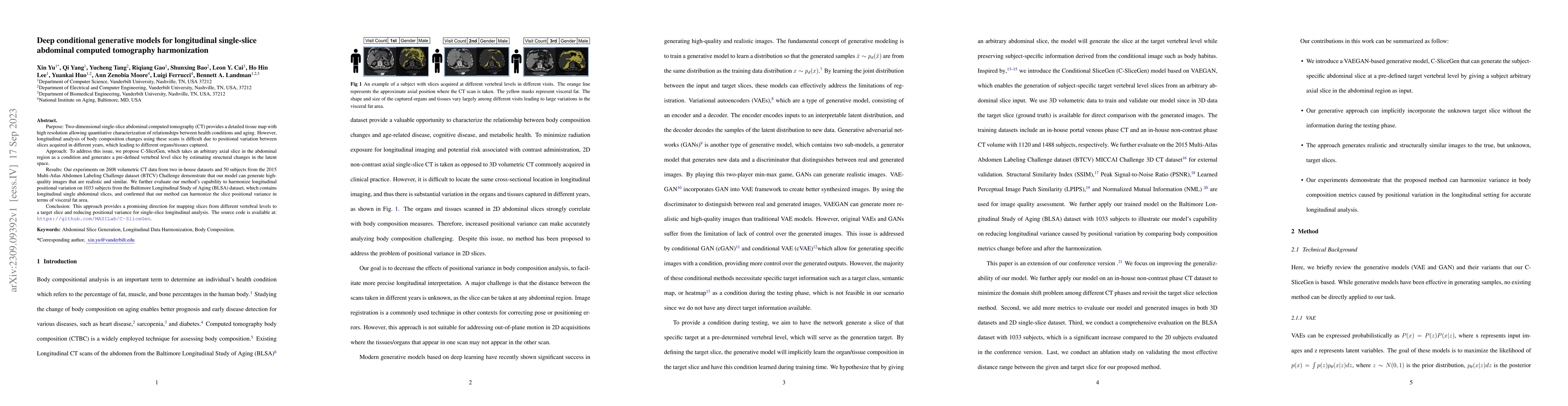
Two-dimensional single-slice abdominal computed tomography (CT) provides a detailed tissue map with high resolution allowing quantitative characterization of relationships between health conditions ...
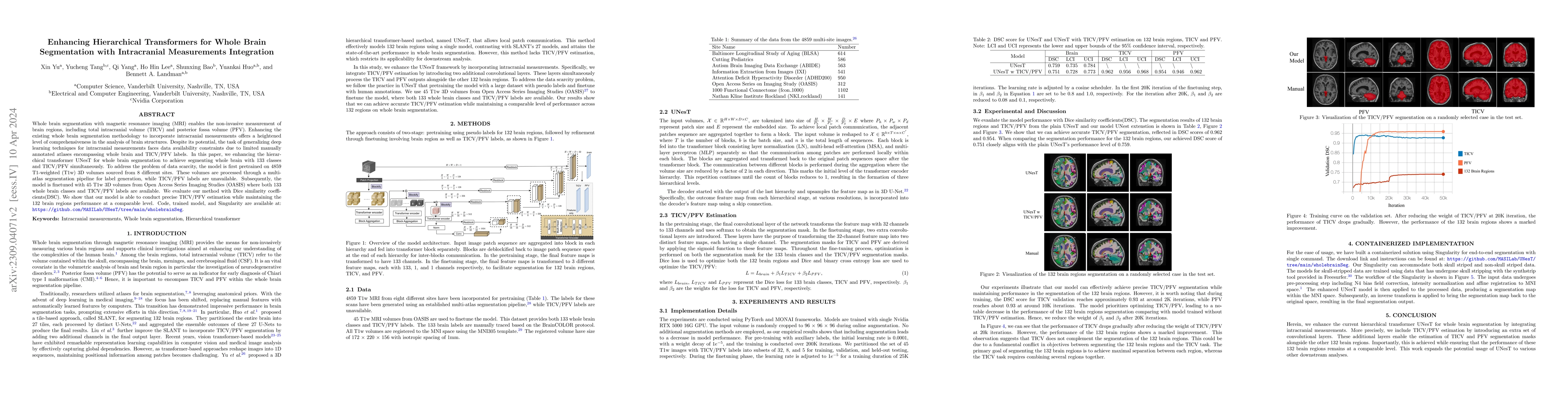
Whole brain segmentation with magnetic resonance imaging (MRI) enables the non-invasive measurement of brain regions, including total intracranial volume (TICV) and posterior fossa volume (PFV). Enh...
The segmentation of kidney layer structures, including cortex, outer stripe, inner stripe, and inner medulla within human kidney whole slide images (WSI) plays an essential role in automated image a...
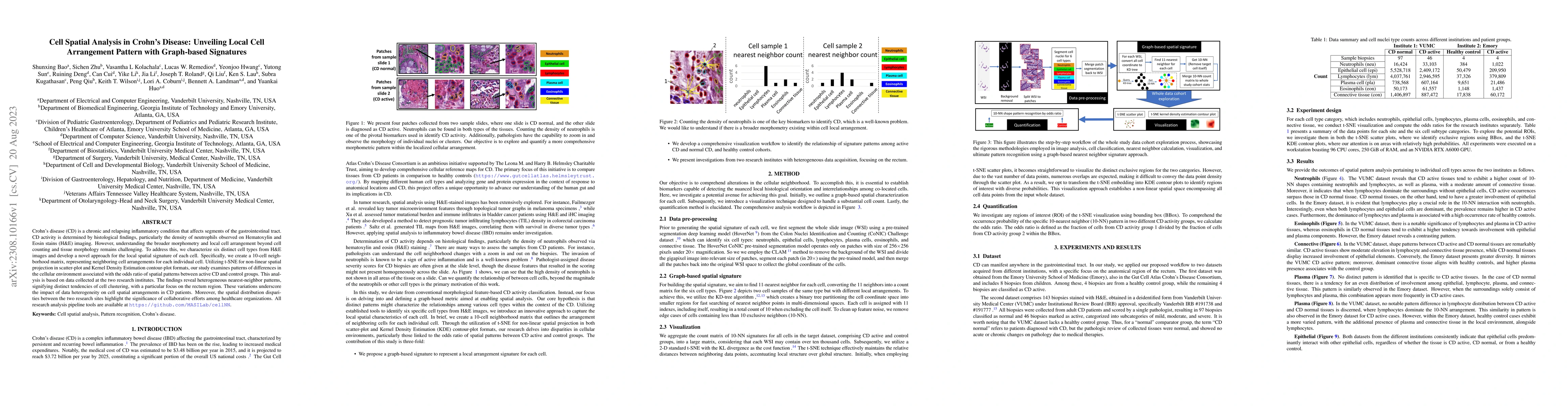
Crohn's disease (CD) is a chronic and relapsing inflammatory condition that affects segments of the gastrointestinal tract. CD activity is determined by histological findings, particularly the densi...
Precise identification of multiple cell classes in high-resolution Giga-pixel whole slide imaging (WSI) is critical for various clinical scenarios. Building an AI model for this purpose typically re...
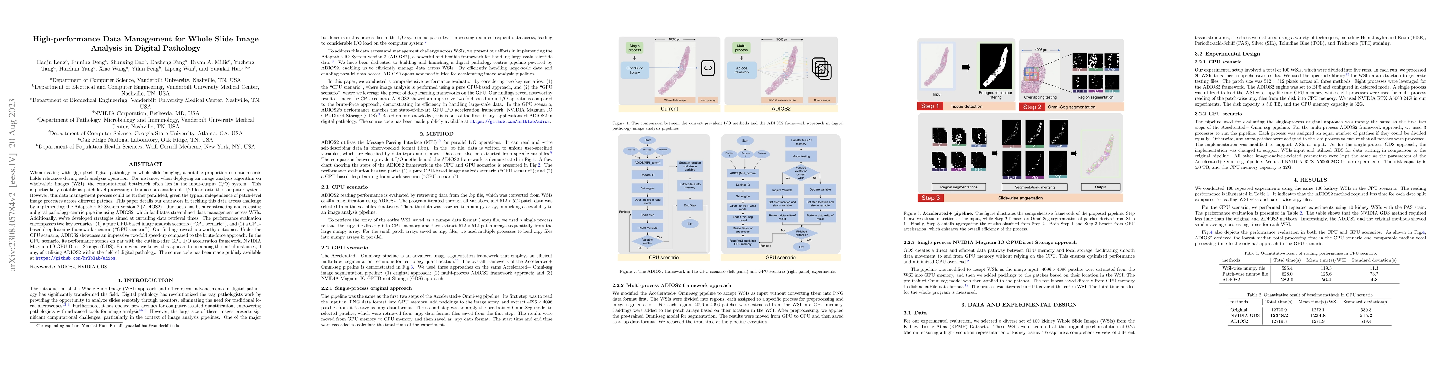
When dealing with giga-pixel digital pathology in whole-slide imaging, a notable proportion of data records holds relevance during each analysis operation. For instance, when deploying an image anal...
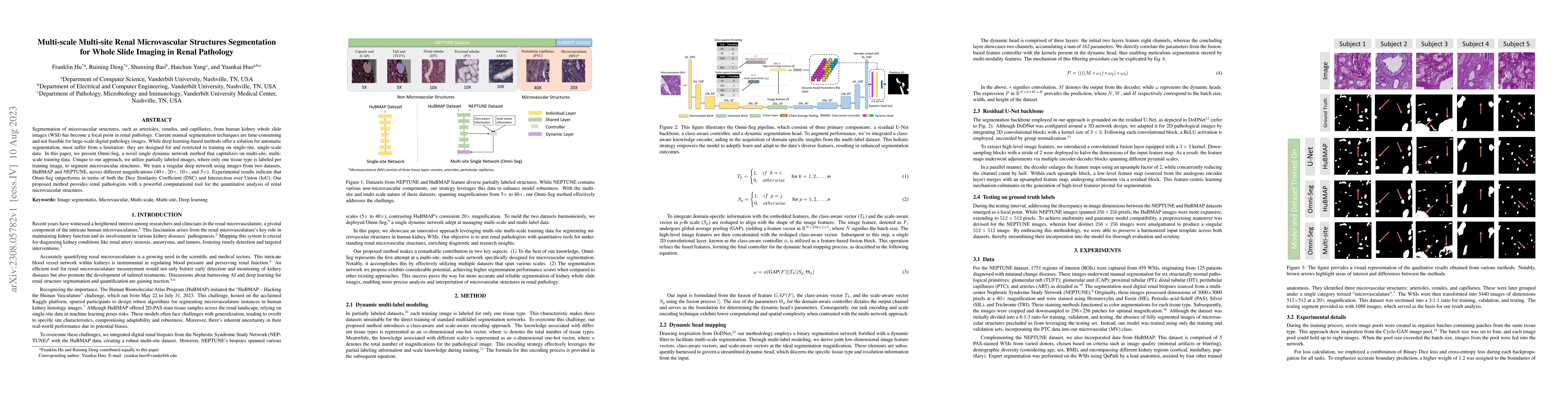
Segmentation of microvascular structures, such as arterioles, venules, and capillaries, from human kidney whole slide images (WSI) has become a focal point in renal pathology. Current manual segment...
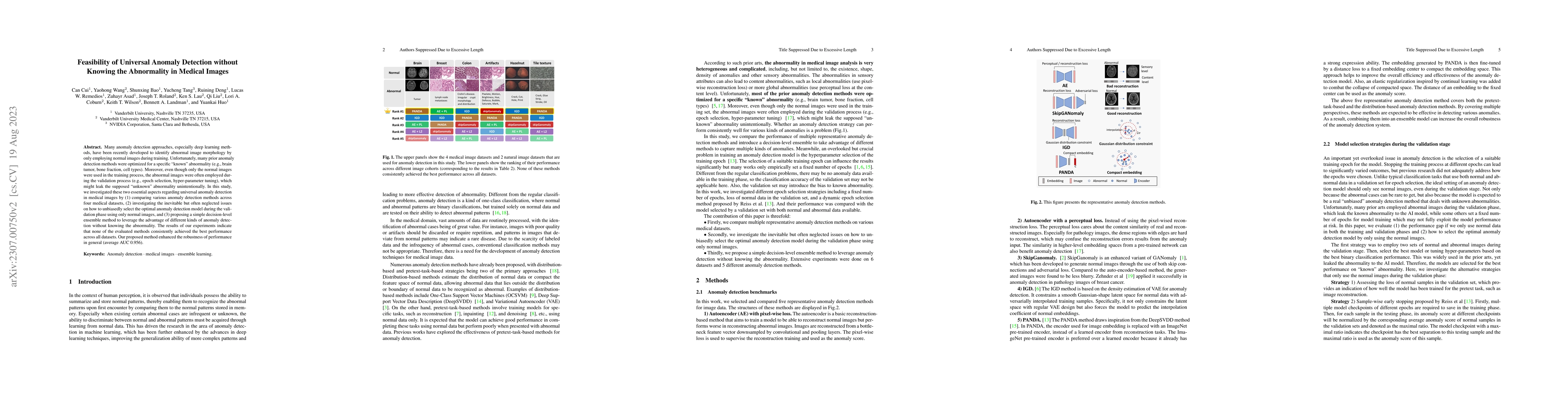
Many anomaly detection approaches, especially deep learning methods, have been recently developed to identify abnormal image morphology by only employing normal images during training. Unfortunately...
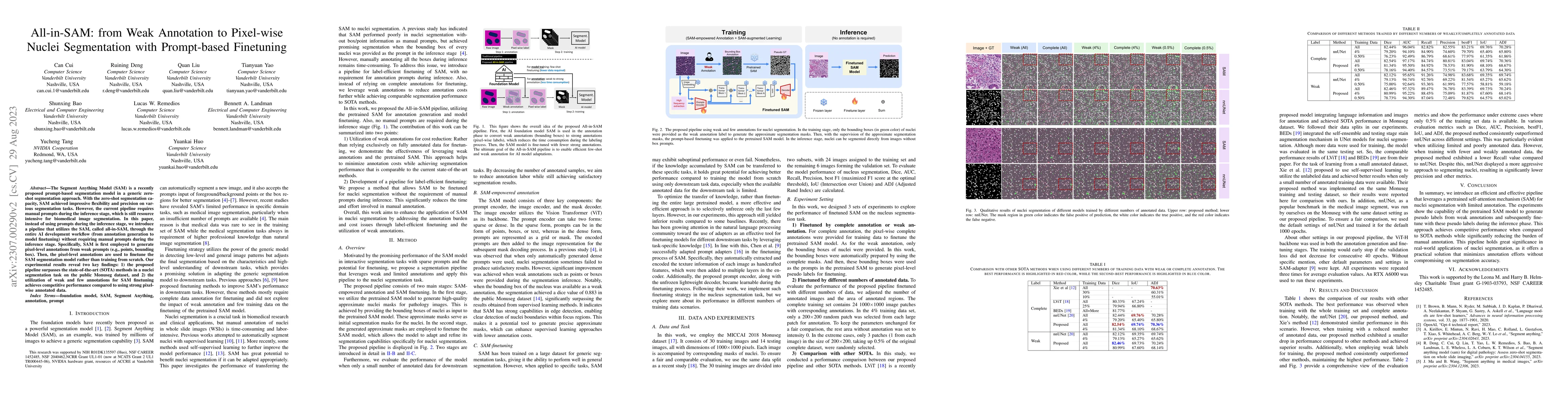
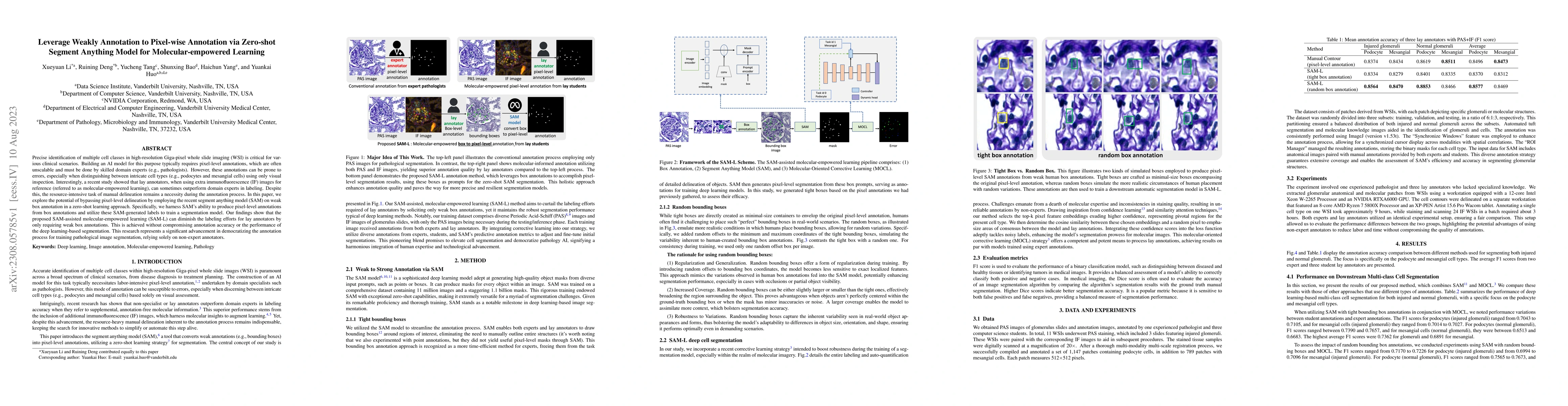
The Segment Anything Model (SAM) is a recently proposed prompt-based segmentation model in a generic zero-shot segmentation approach. With the zero-shot segmentation capacity, SAM achieved impressiv...
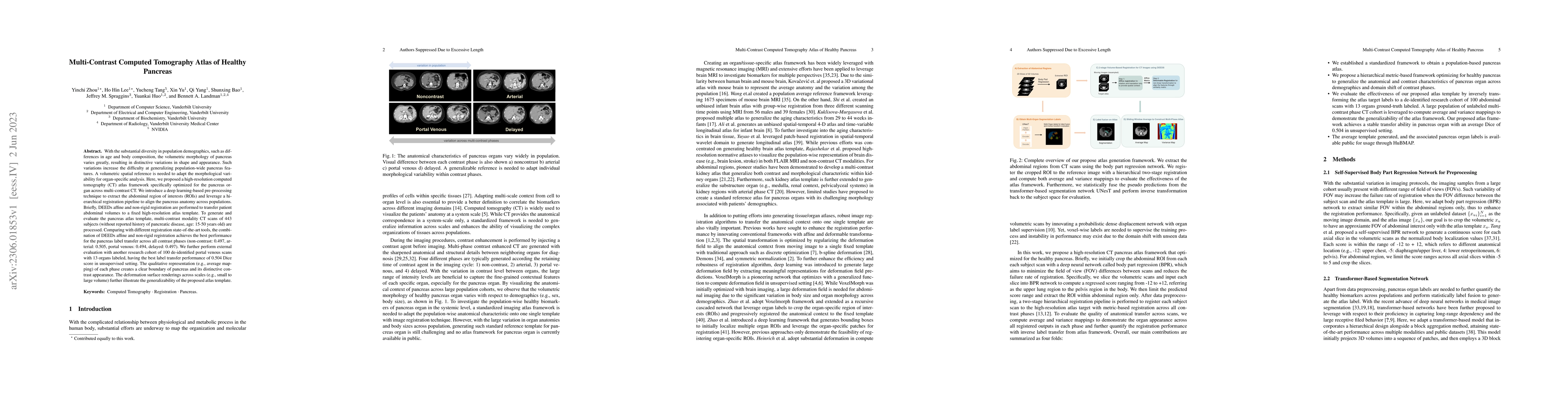
With the substantial diversity in population demographics, such as differences in age and body composition, the volumetric morphology of pancreas varies greatly, resulting in distinctive variations ...
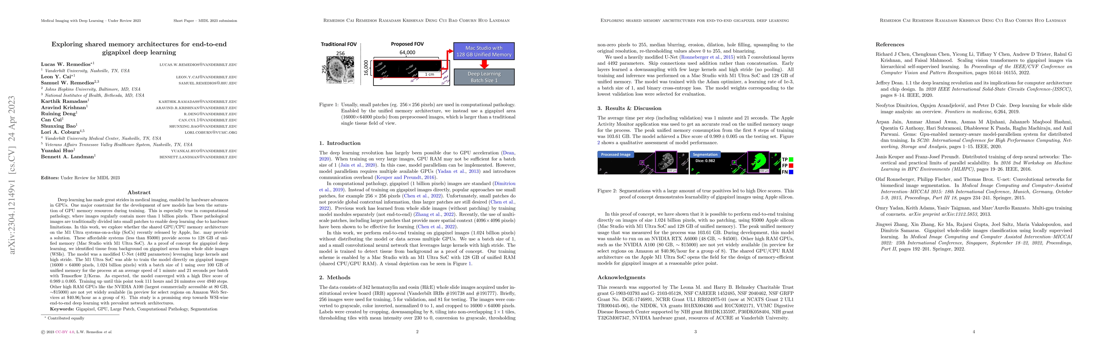
Deep learning has made great strides in medical imaging, enabled by hardware advances in GPUs. One major constraint for the development of new models has been the saturation of GPU memory resources ...
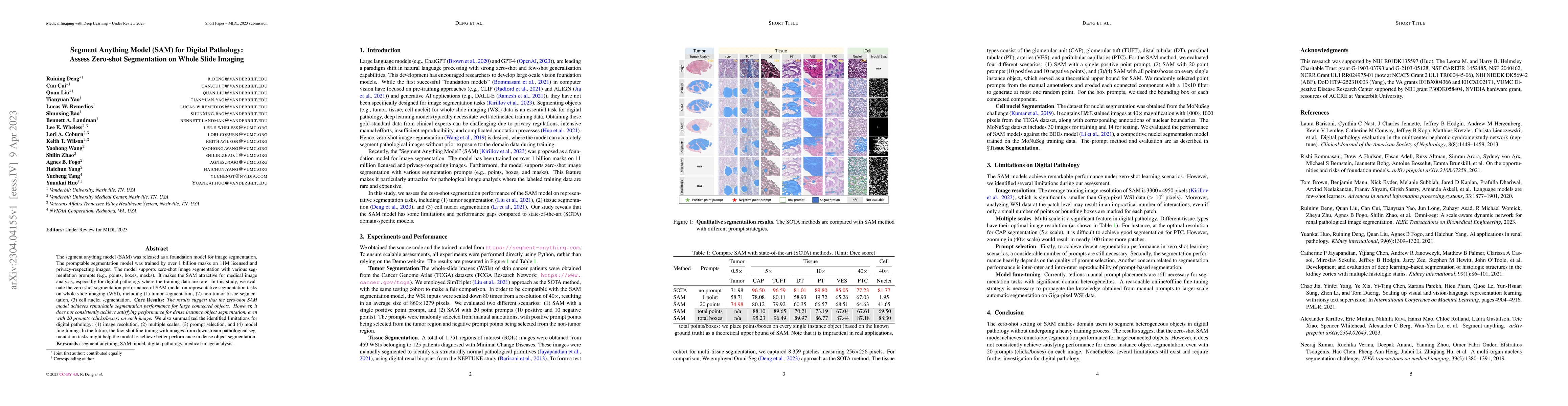
The segment anything model (SAM) was released as a foundation model for image segmentation. The promptable segmentation model was trained by over 1 billion masks on 11M licensed and privacy-respecti...
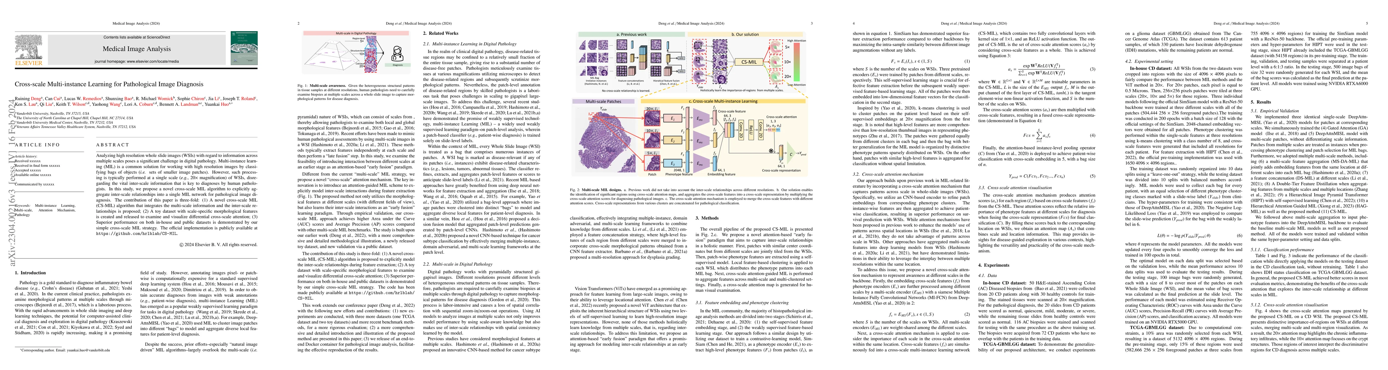
Analyzing high resolution whole slide images (WSIs) with regard to information across multiple scales poses a significant challenge in digital pathology. Multi-instance learning (MIL) is a common so...
With the inspiration of vision transformers, the concept of depth-wise convolution revisits to provide a large Effective Receptive Field (ERF) using Large Kernel (LK) sizes for medical image segment...
The Distributed Messaging Systems (DMSs) used in IoT systems require timely and reliable data dissemination, which can be achieved through configurable parameters. However, the high-dimensional conf...
Objective: Thigh muscle group segmentation is important for assessment of muscle anatomy, metabolic disease and aging. Many efforts have been put into quantifying muscle tissues with magnetic resona...
Recent studies have demonstrated the superior performance of introducing ``scan-wise" contrast labels into contrastive learning for multi-organ segmentation on multi-phase computed tomography (CT). ...
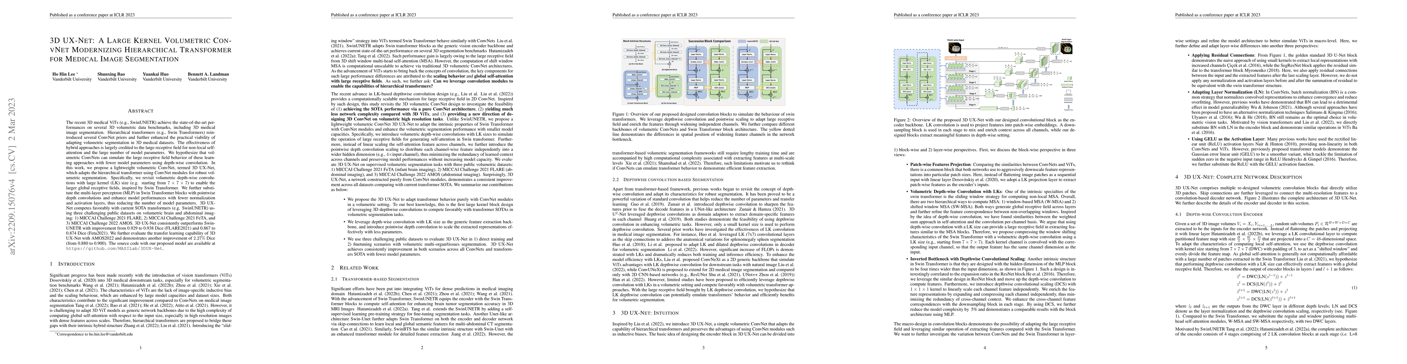
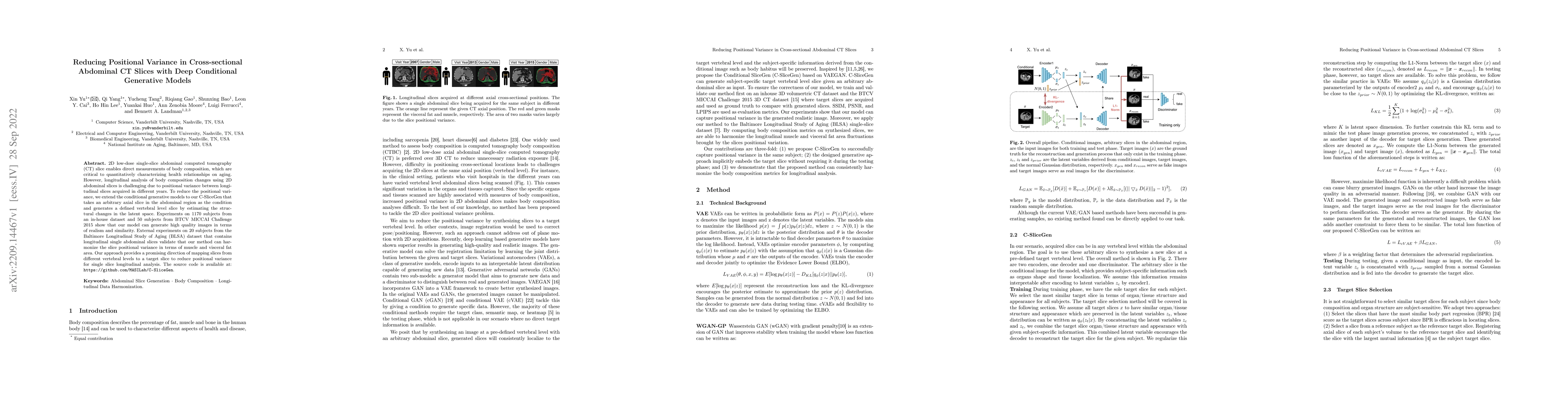
The recent 3D medical ViTs (e.g., SwinUNETR) achieve the state-of-the-art performances on several 3D volumetric data benchmarks, including 3D medical image segmentation. Hierarchical transformers (e...
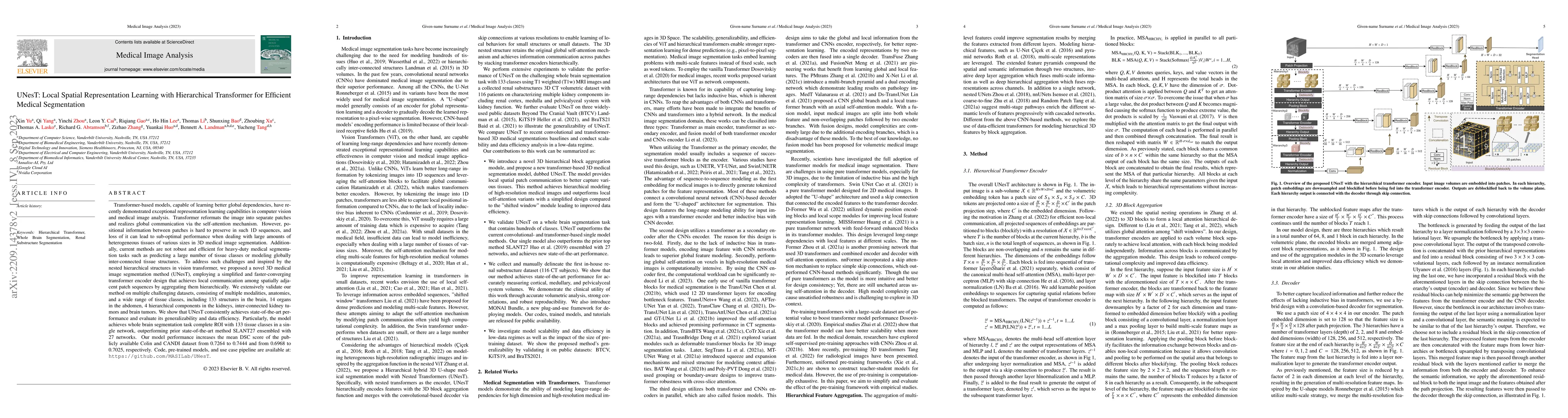
2D low-dose single-slice abdominal computed tomography (CT) slice enables direct measurements of body composition, which are critical to quantitatively characterizing health relationships on aging. ...
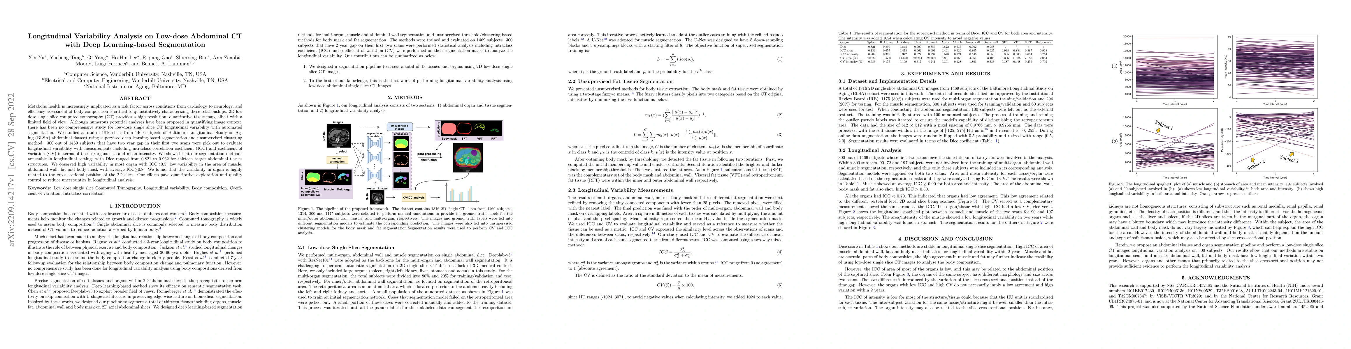
Transformer-based models, capable of learning better global dependencies, have recently demonstrated exceptional representation learning capabilities in computer vision and medical image analysis. T...
Metabolic health is increasingly implicated as a risk factor across conditions from cardiology to neurology, and efficiency assessment of body composition is critical to quantitatively characterizin...
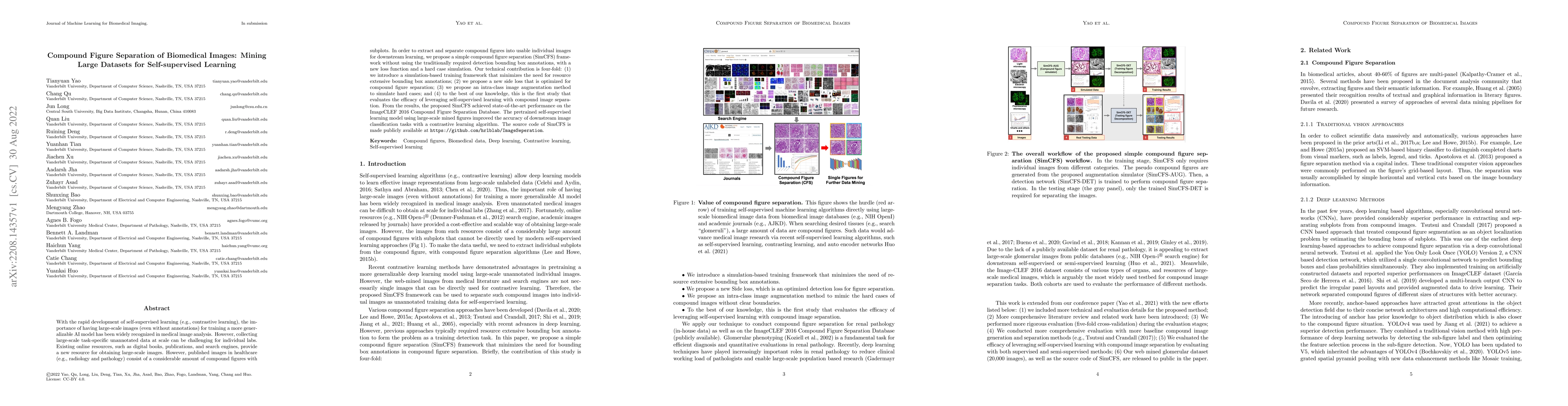
With the rapid development of self-supervised learning (e.g., contrastive learning), the importance of having large-scale images (even without annotations) for training a more generalizable AI model...
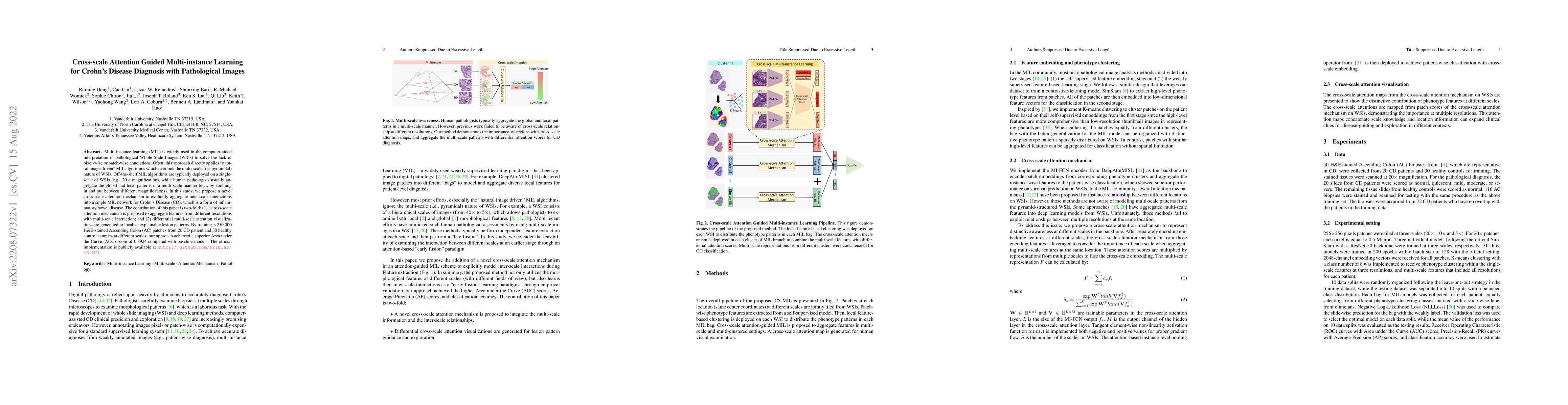
Multi-instance learning (MIL) is widely used in the computer-aided interpretation of pathological Whole Slide Images (WSIs) to solve the lack of pixel-wise or patch-wise annotations. Often, this app...
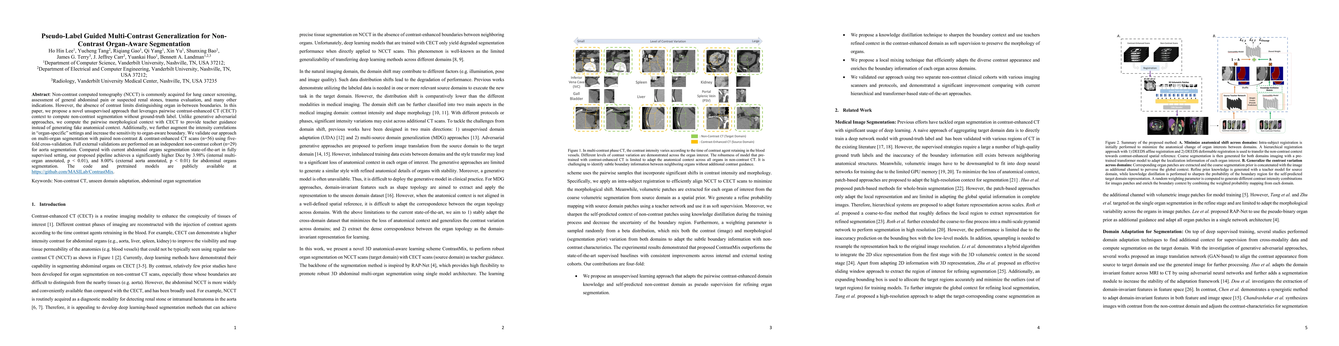
Non-contrast computed tomography (NCCT) is commonly acquired for lung cancer screening, assessment of general abdominal pain or suspected renal stones, trauma evaluation, and many other indications....
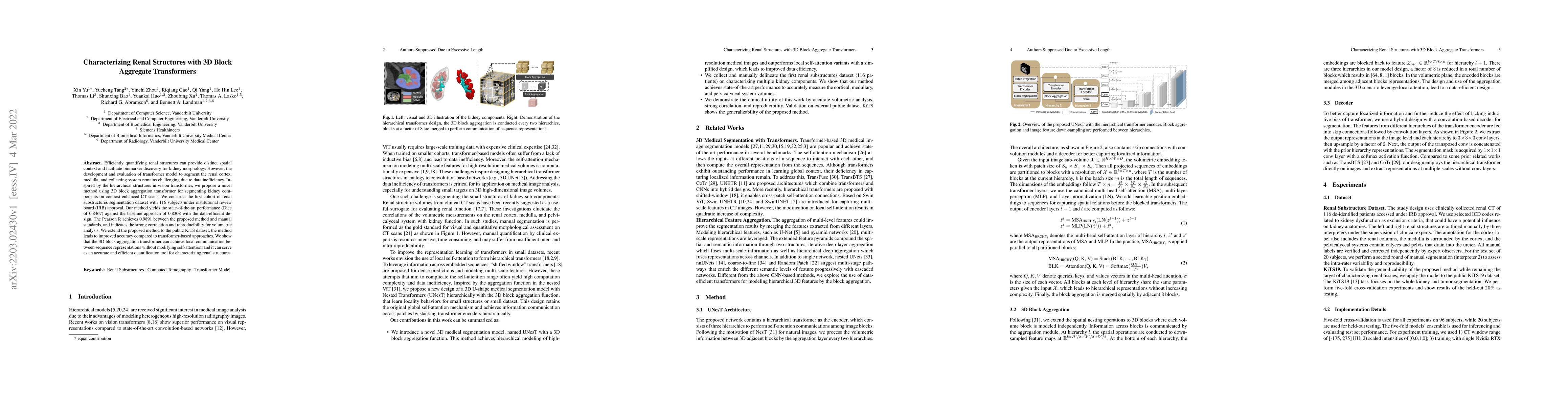
Efficiently quantifying renal structures can provide distinct spatial context and facilitate biomarker discovery for kidney morphology. However, the development and evaluation of the transformer mod...
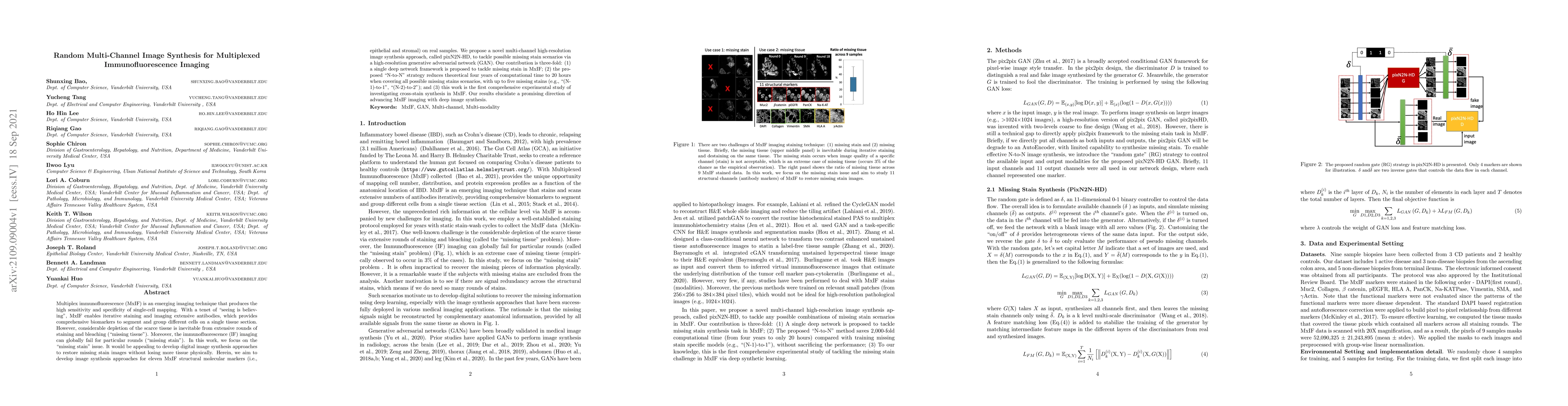
Multiplex immunofluorescence (MxIF) is an emerging imaging technique that produces the high sensitivity and specificity of single-cell mapping. With a tenet of 'seeing is believing', MxIF enables it...
Unsupervised learning algorithms (e.g., self-supervised learning, auto-encoder, contrastive learning) allow deep learning models to learn effective image representations from large-scale unlabeled d...

Medical image segmentation, or computing voxelwise semantic masks, is a fundamental yet challenging task to compute a voxel-level semantic mask. To increase the ability of encoder-decoder neural net...
The construction of three-dimensional multi-modal tissue maps provides an opportunity to spur interdisciplinary innovations across temporal and spatial scales through information integration. While ...
A major goal of lung cancer screening is to identify individuals with particular phenotypes that are associated with high risk of cancer. Identifying relevant phenotypes is complicated by the variat...
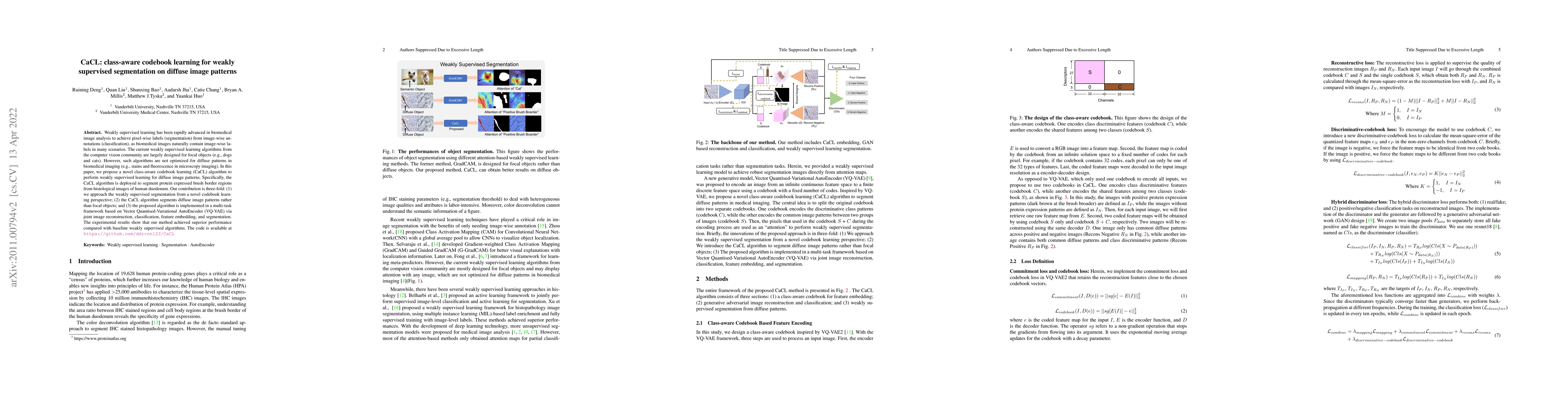
Weakly supervised learning has been rapidly advanced in biomedical image analysis to achieve pixel-wise labels (segmentation) from image-wise annotations (classification), as biomedical images natur...
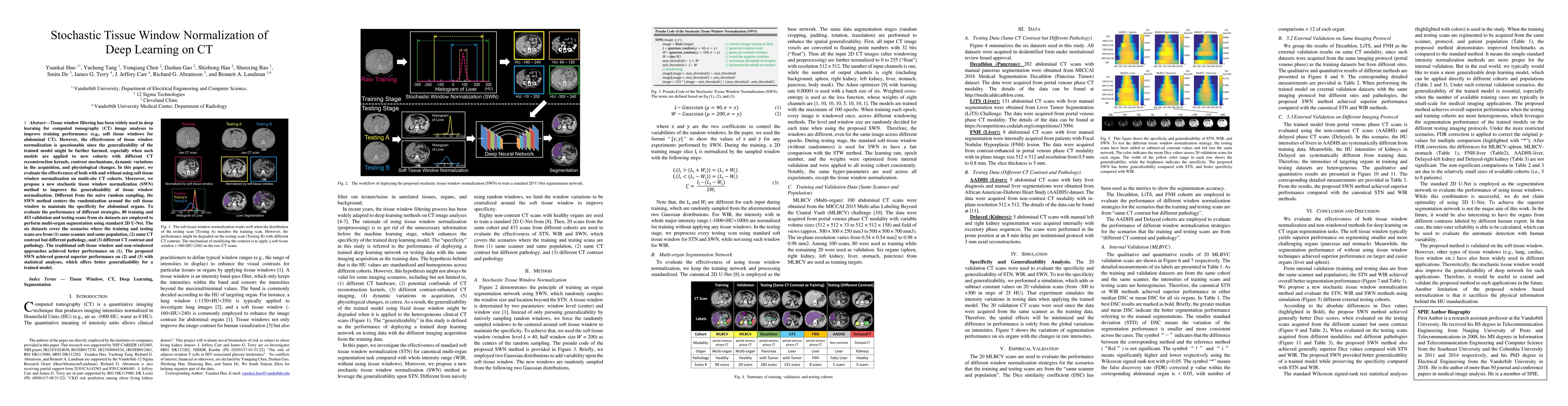
Tissue window filtering has been widely used in deep learning for computed tomography (CT) image analyses to improve training performance (e.g., soft tissue windows for abdominal CT). However, the e...
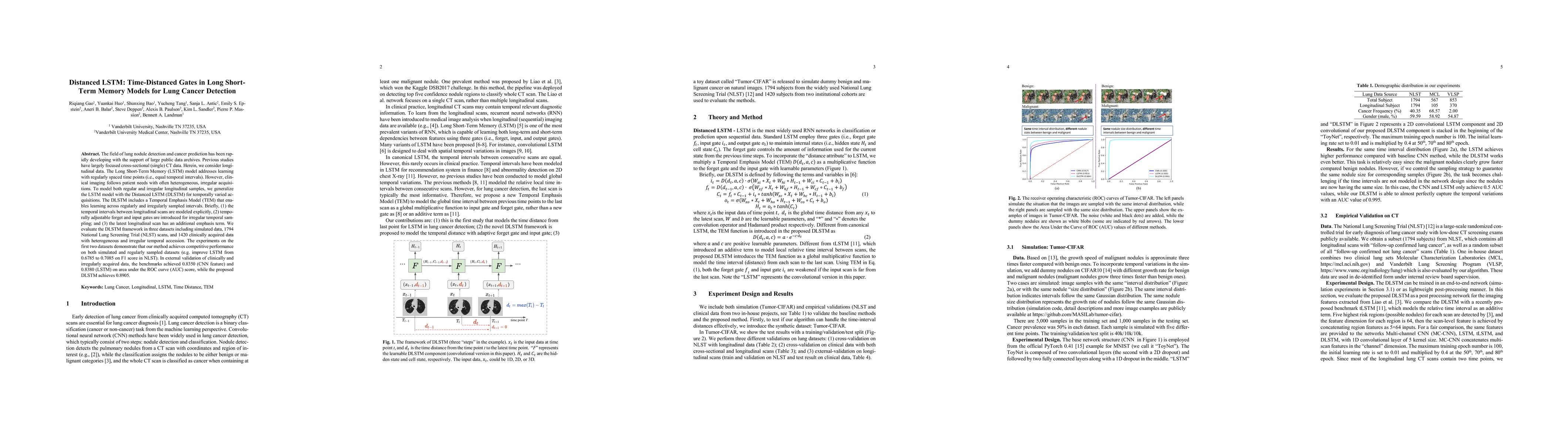
The field of lung nodule detection and cancer prediction has been rapidly developing with the support of large public data archives. Previous studies have largely focused on cross-sectional (single)...
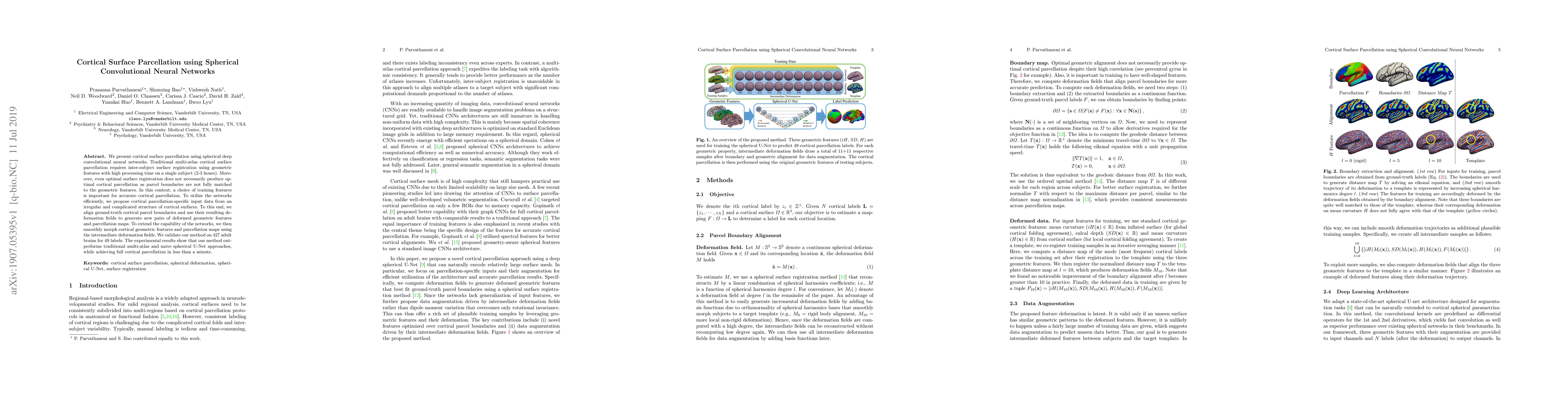
We present cortical surface parcellation using spherical deep convolutional neural networks. Traditional multi-atlas cortical surface parcellation requires inter-subject surface registration using g...
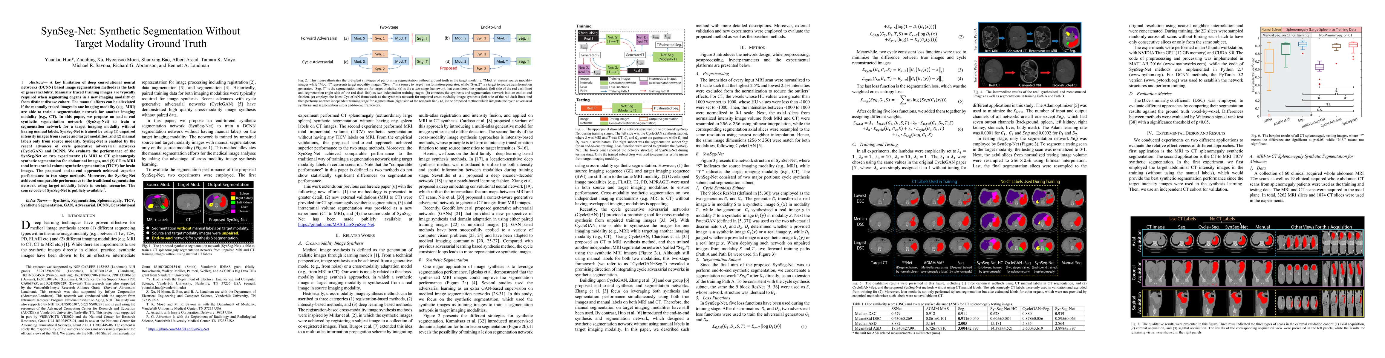
A key limitation of deep convolutional neural networks (DCNN) based image segmentation methods is the lack of generalizability. Manually traced training images are typically required when segmenting...
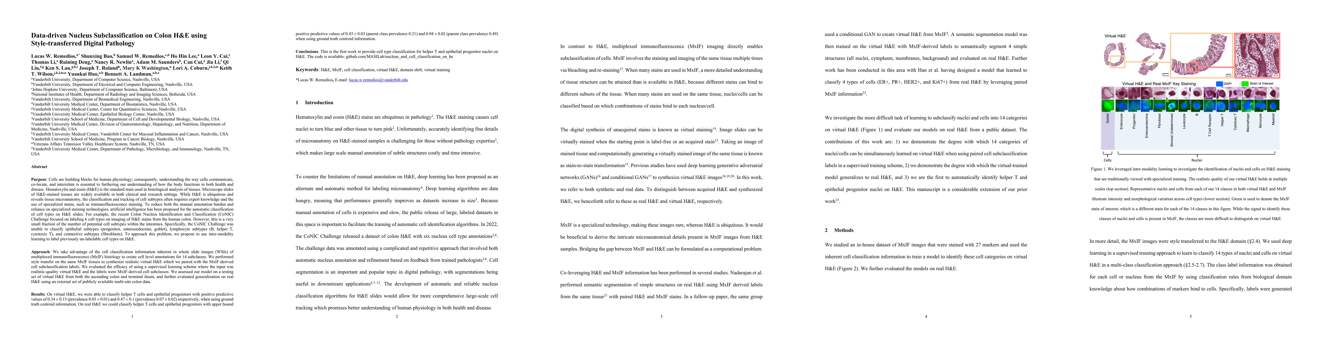
Understanding the way cells communicate, co-locate, and interrelate is essential to furthering our understanding of how the body functions. H&E is widely available, however, cell subtyping often requi...
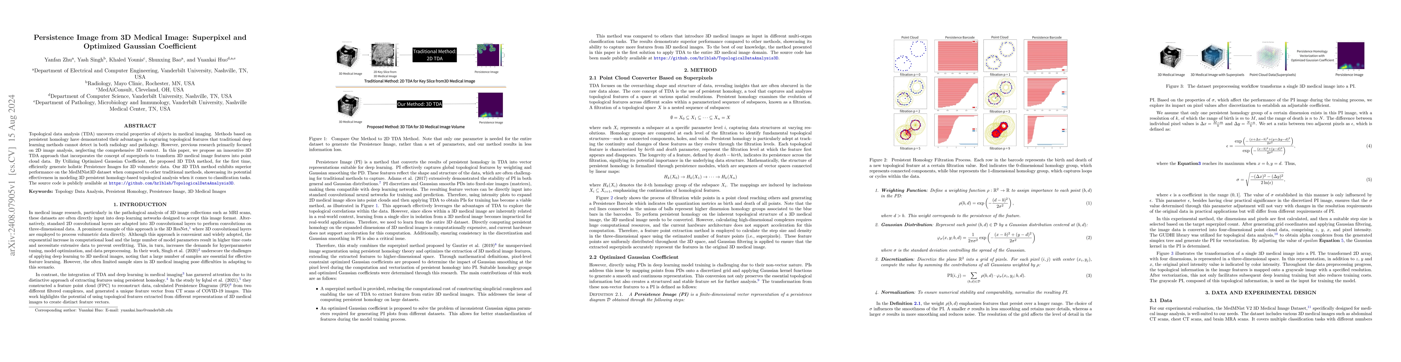
Topological data analysis (TDA) uncovers crucial properties of objects in medical imaging. Methods based on persistent homology have demonstrated their advantages in capturing topological features tha...
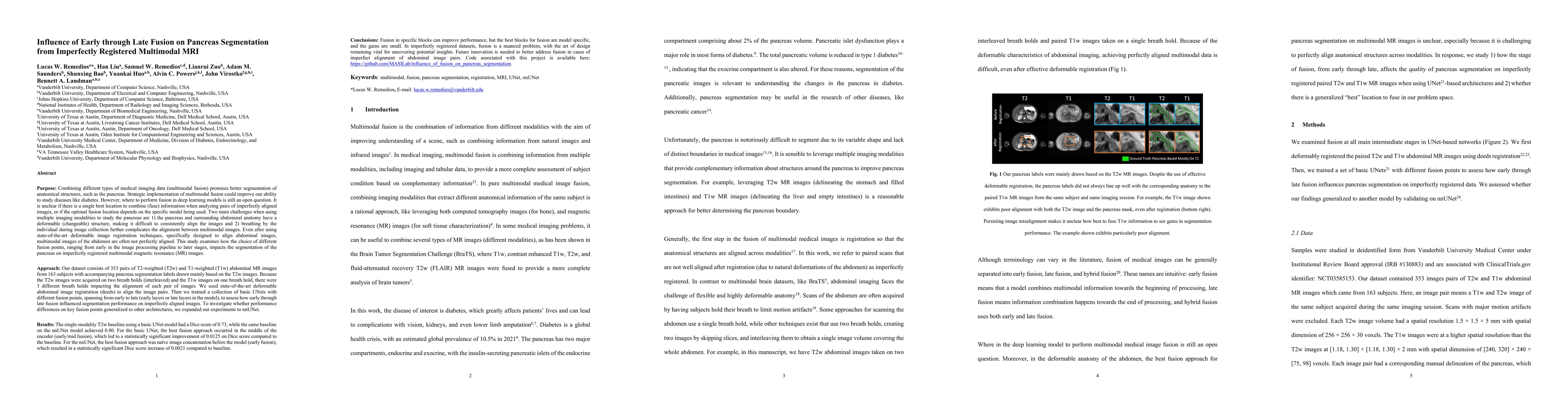
Multimodal fusion promises better pancreas segmentation. However, where to perform fusion in models is still an open question. It is unclear if there is a best location to fuse information when analyz...
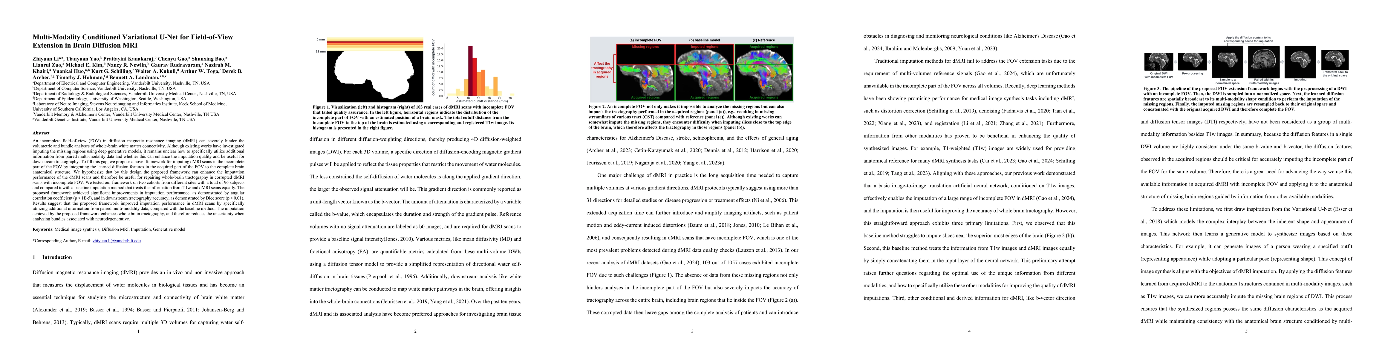
An incomplete field-of-view (FOV) in diffusion magnetic resonance imaging (dMRI) can severely hinder the volumetric and bundle analyses of whole-brain white matter connectivity. Although existing work...
Estimated brain age from magnetic resonance image (MRI) and its deviation from chronological age can provide early insights into potential neurodegenerative diseases, supporting early detection and im...
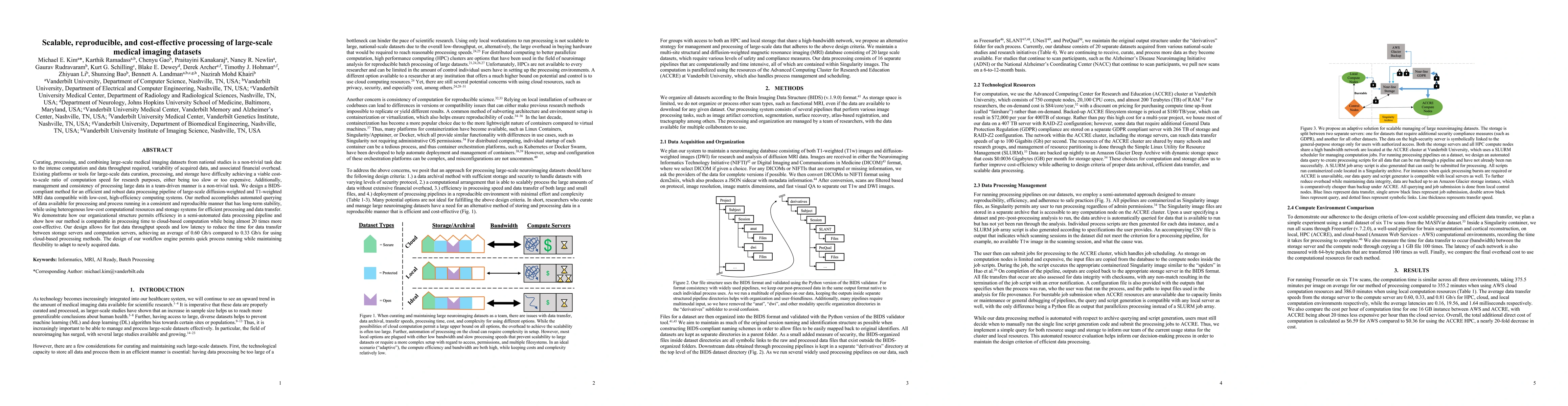
Curating, processing, and combining large-scale medical imaging datasets from national studies is a non-trivial task due to the intense computation and data throughput required, variability of acquire...
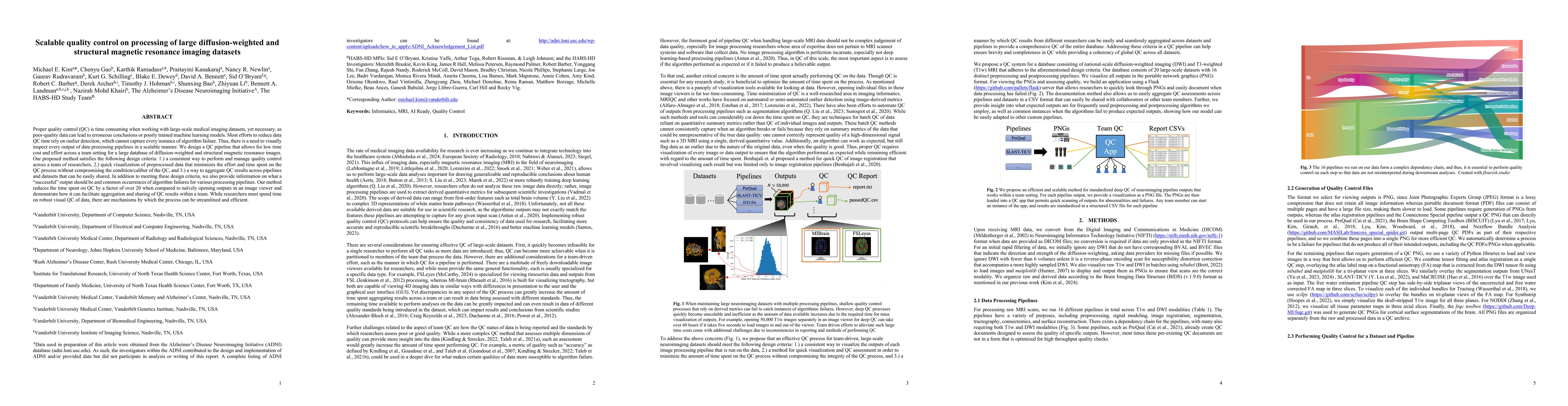
Proper quality control (QC) is time consuming when working with large-scale medical imaging datasets, yet necessary, as poor-quality data can lead to erroneous conclusions or poorly trained machine le...
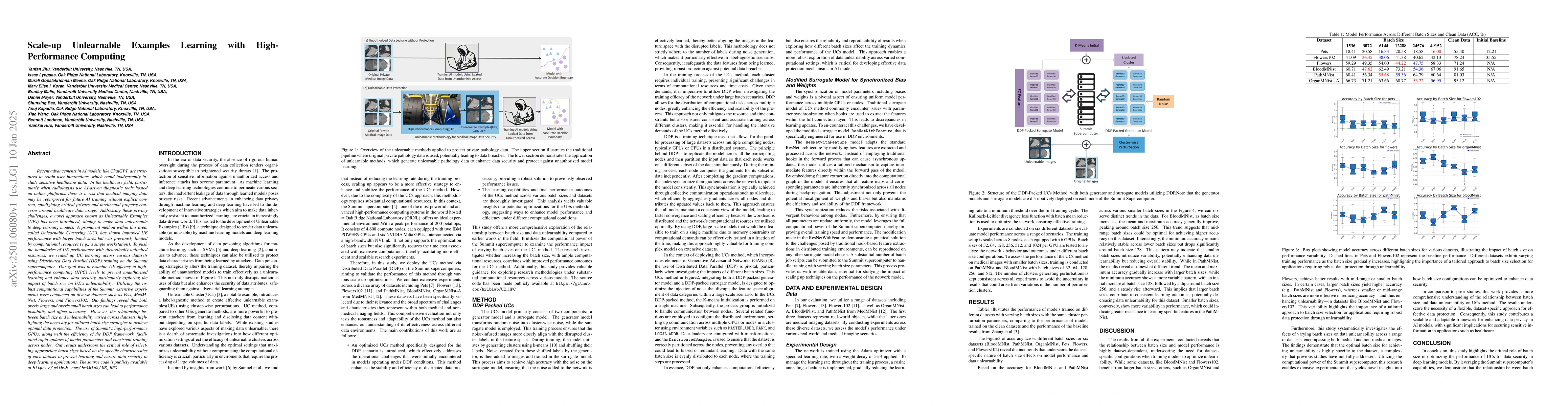
Recent advancements in AI models are structured to retain user interactions, which could inadvertently include sensitive healthcare data. In the healthcare field, particularly when radiologists use AI...
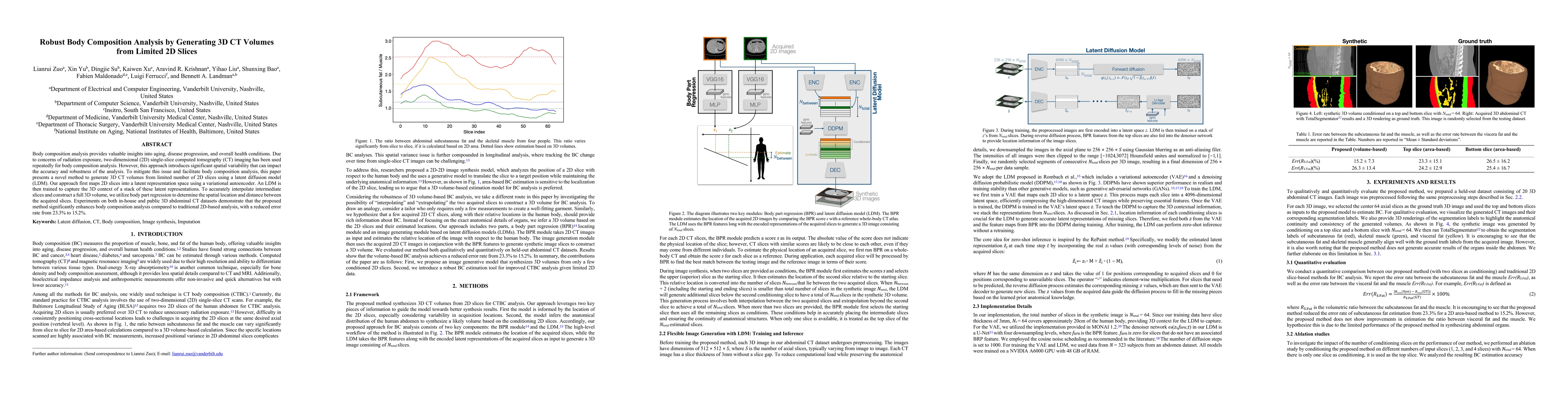
Body composition analysis provides valuable insights into aging, disease progression, and overall health conditions. Due to concerns of radiation exposure, two-dimensional (2D) single-slice computed t...
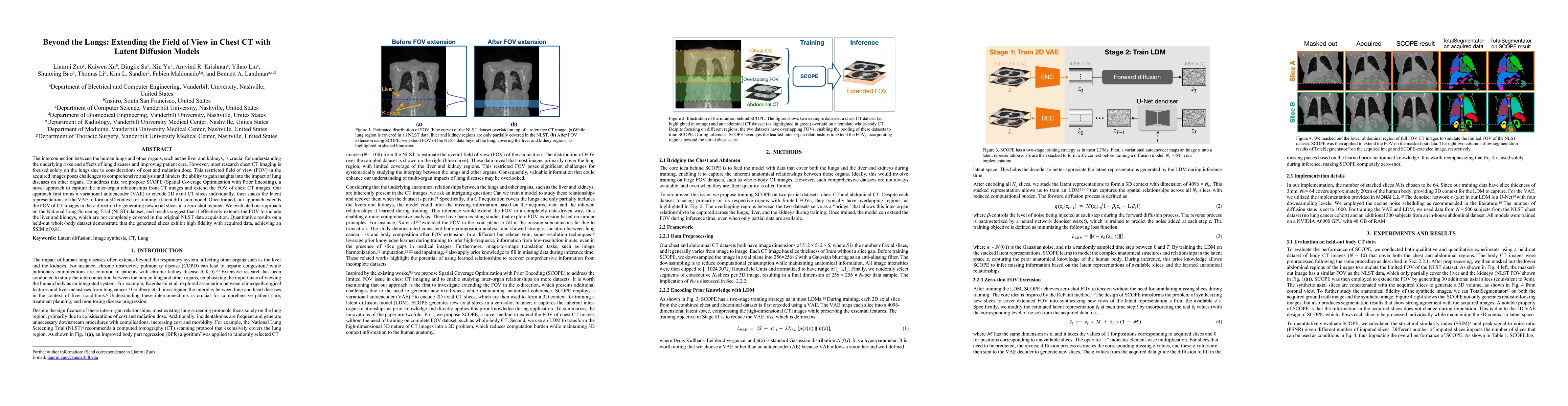
The interconnection between the human lungs and other organs, such as the liver and kidneys, is crucial for understanding the underlying risks and effects of lung diseases and improving patient care. ...
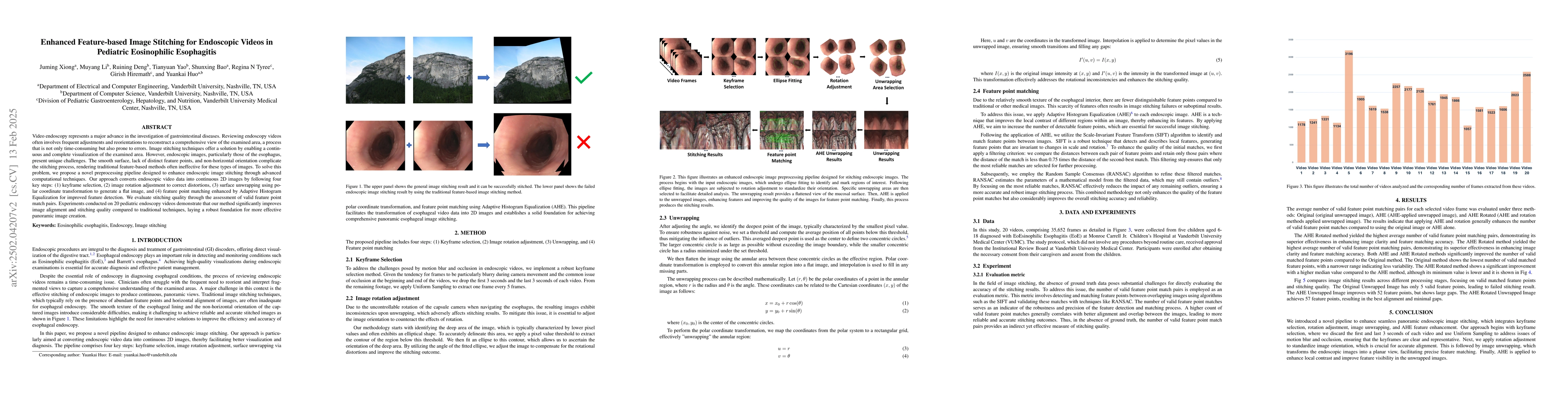
Video endoscopy represents a major advance in the investigation of gastrointestinal diseases. Reviewing endoscopy videos often involves frequent adjustments and reorientations to piece together a comp...
Reconstruction kernels in computed tomography (CT) affect spatial resolution and noise characteristics, introducing systematic variability in quantitative imaging measurements such as emphysema quanti...
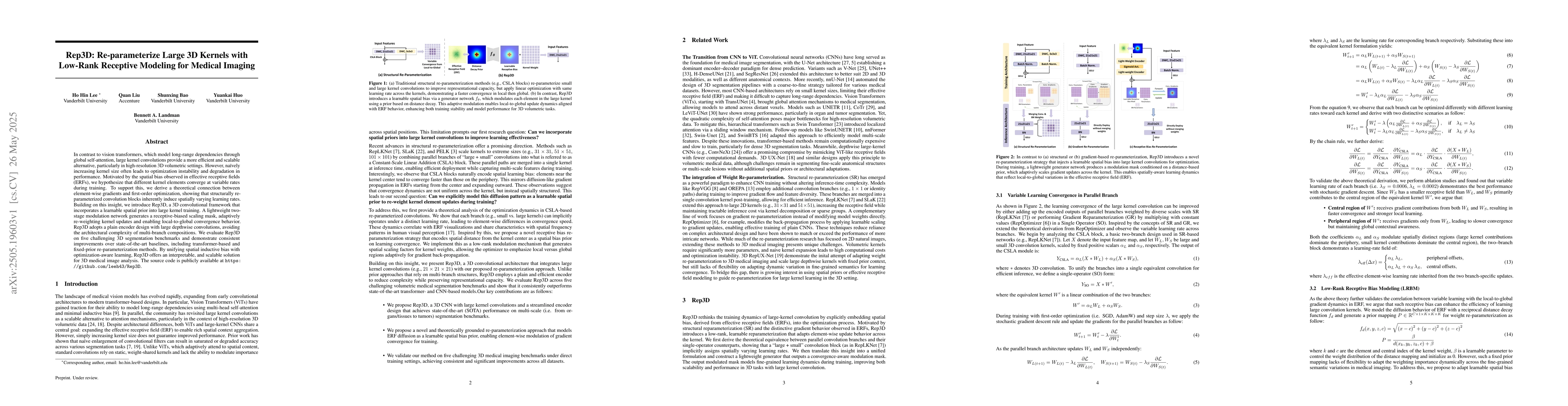
In contrast to vision transformers, which model long-range dependencies through global self-attention, large kernel convolutions provide a more efficient and scalable alternative, particularly in high...
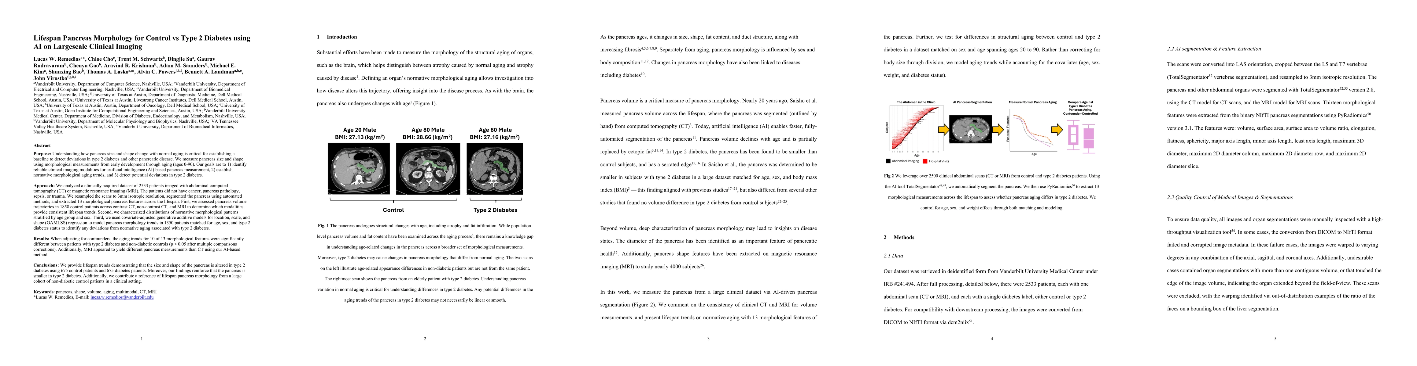
Purpose: Understanding how the pancreas changes is critical for detecting deviations in type 2 diabetes and other pancreatic disease. We measure pancreas size and shape using morphological measurement...
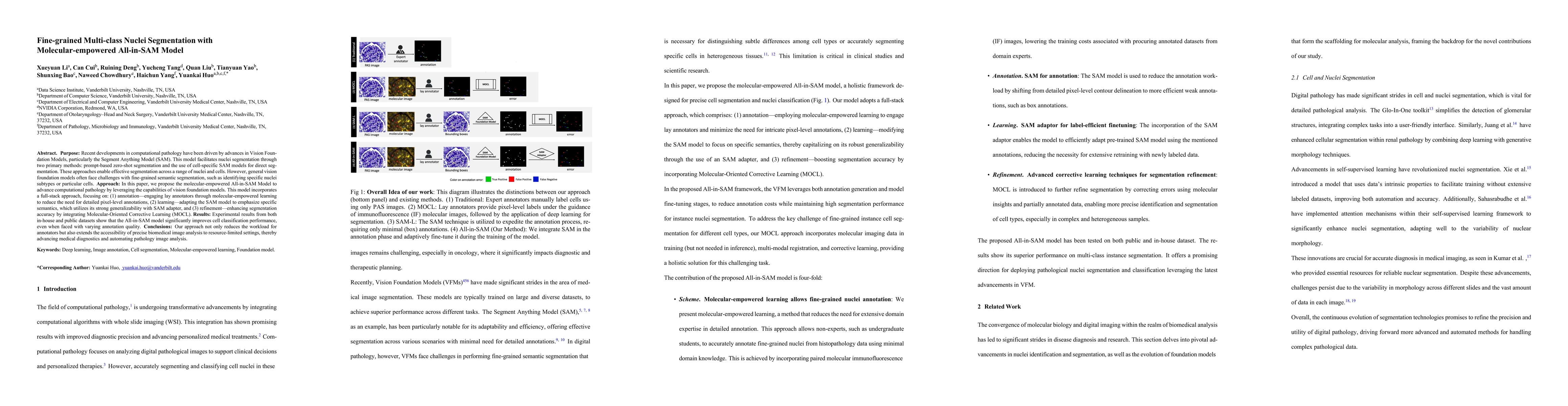
Purpose: Recent developments in computational pathology have been driven by advances in Vision Foundation Models, particularly the Segment Anything Model (SAM). This model facilitates nuclei segmentat...
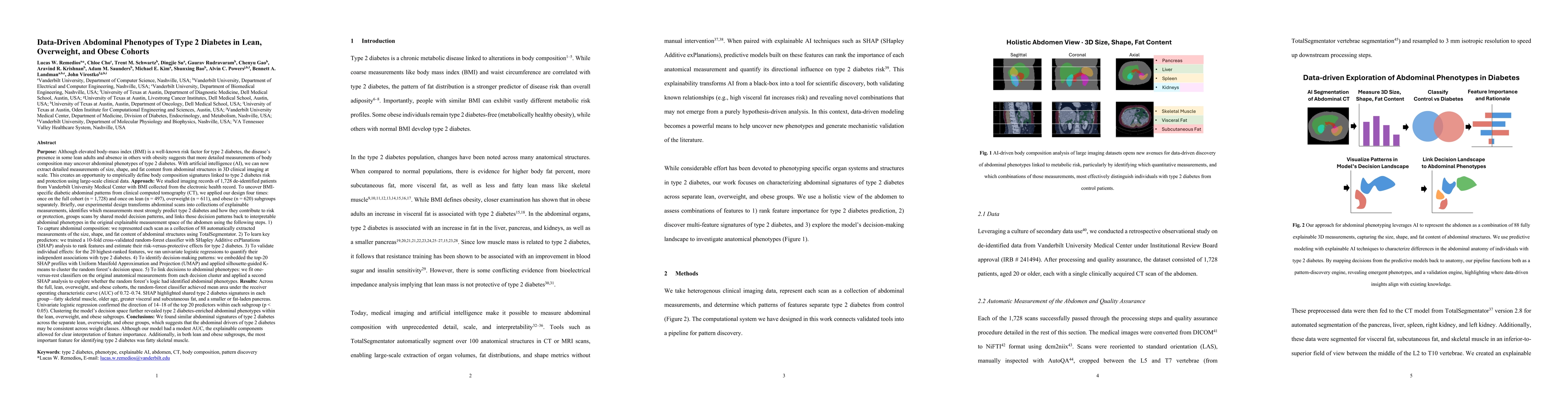
Purpose: Although elevated BMI is a well-known risk factor for type 2 diabetes, the disease's presence in some lean adults and absence in others with obesity suggests that detailed body composition ma...