Academic Profile
Statistics
Similar Authors
Papers on arXiv
Functional Magnetic Resonance Imaging (fMRI) is vital in neuroscience, enabling investigations into brain disorders, treatment monitoring, and brain function mapping. However, head motion during fMR...
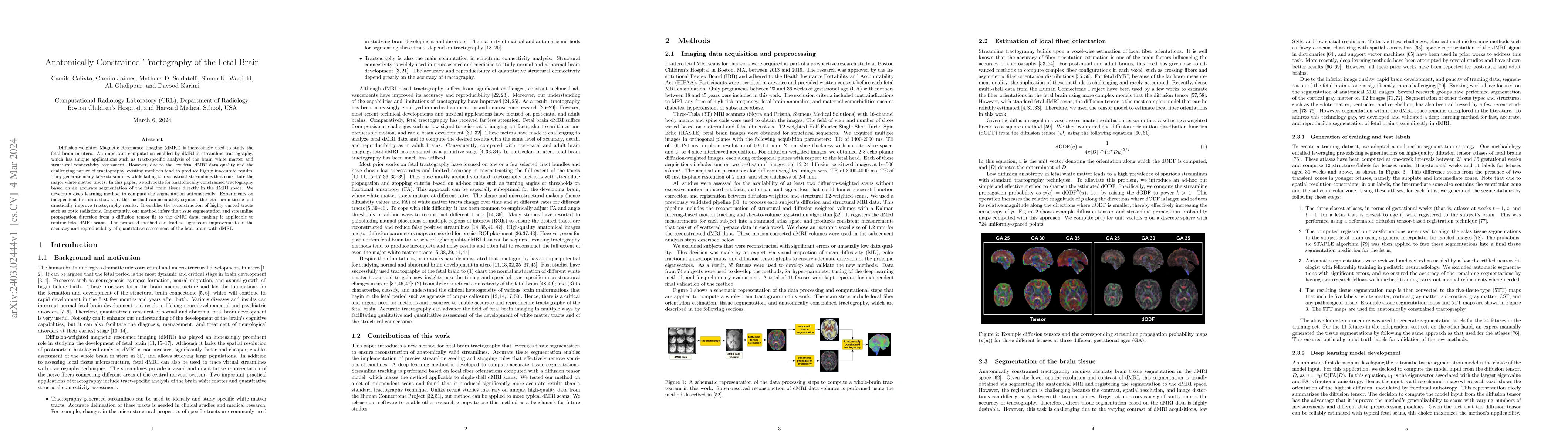
Diffusion-weighted Magnetic Resonance Imaging (dMRI) is increasingly used to study the fetal brain in utero. An important computation enabled by dMRI is streamline tractography, which has unique app...
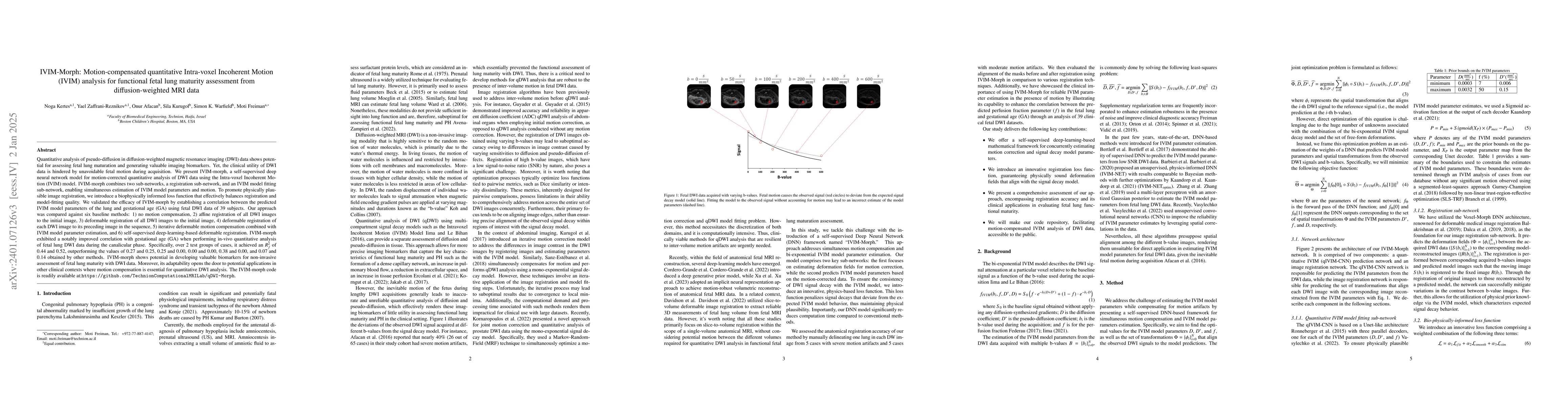
Quantitative analysis of pseudo-diffusion in diffusion-weighted magnetic resonance imaging (DWI) data shows potential for assessing fetal lung maturation and generating valuable imaging biomarkers. ...
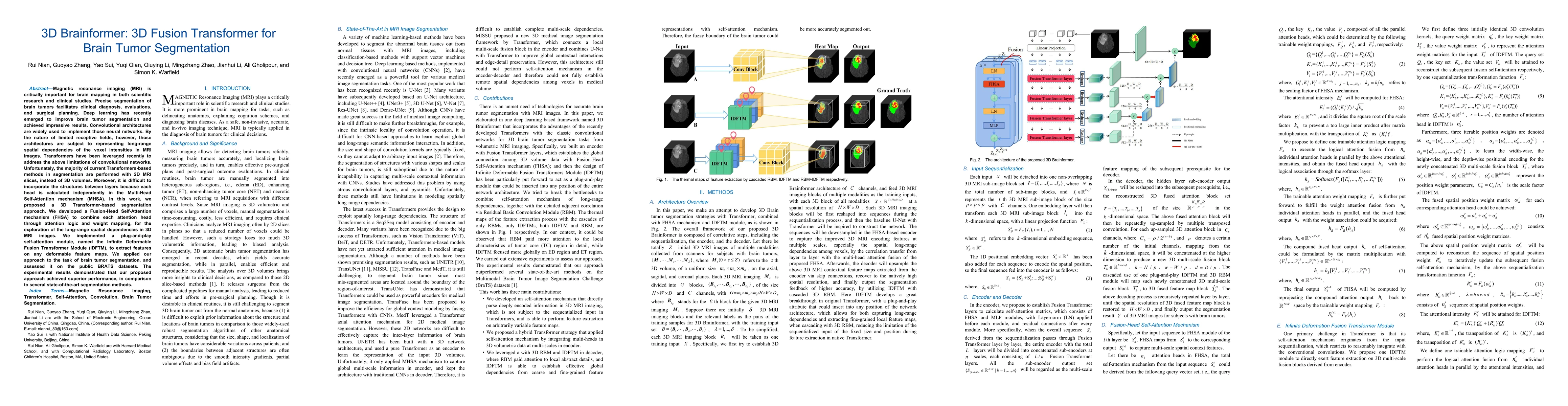
Magnetic resonance imaging (MRI) is critically important for brain mapping in both scientific research and clinical studies. Precise segmentation of brain tumors facilitates clinical diagnosis, eval...
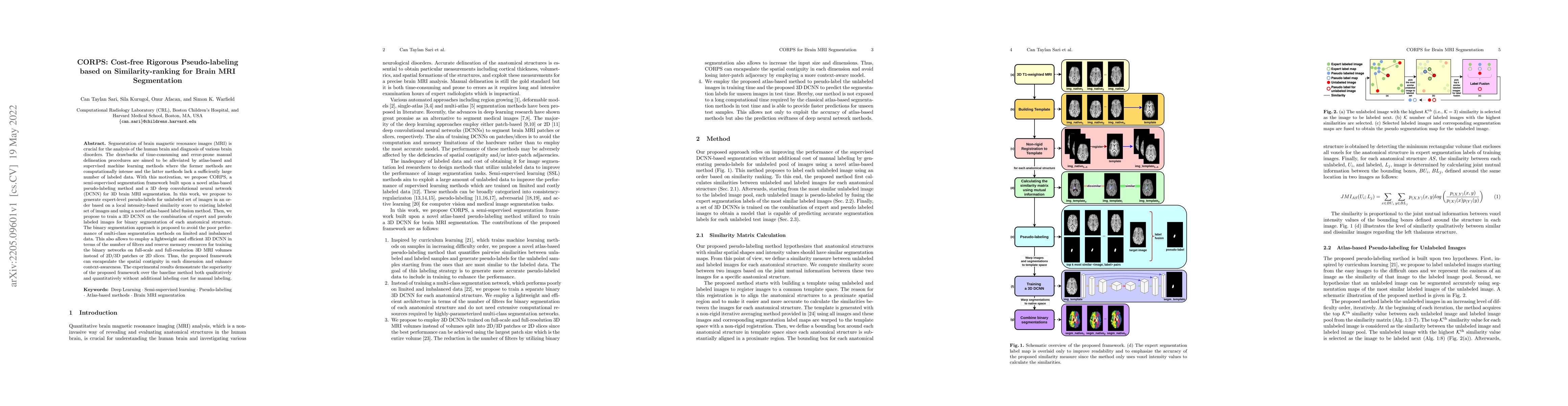
Segmentation of brain magnetic resonance images (MRI) is crucial for the analysis of the human brain and diagnosis of various brain disorders. The drawbacks of time-consuming and error-prone manual ...
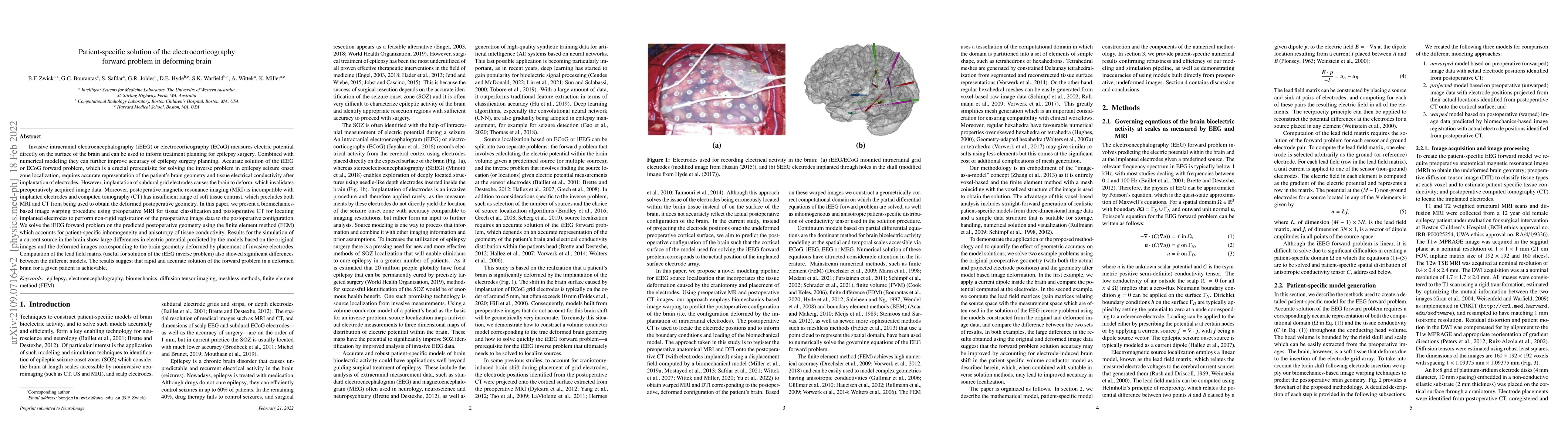
Invasive intracranial electroencephalography (iEEG) or electrocorticography (ECoG) measures electric potential directly on the surface of the brain and can be used to inform treatment planning for e...
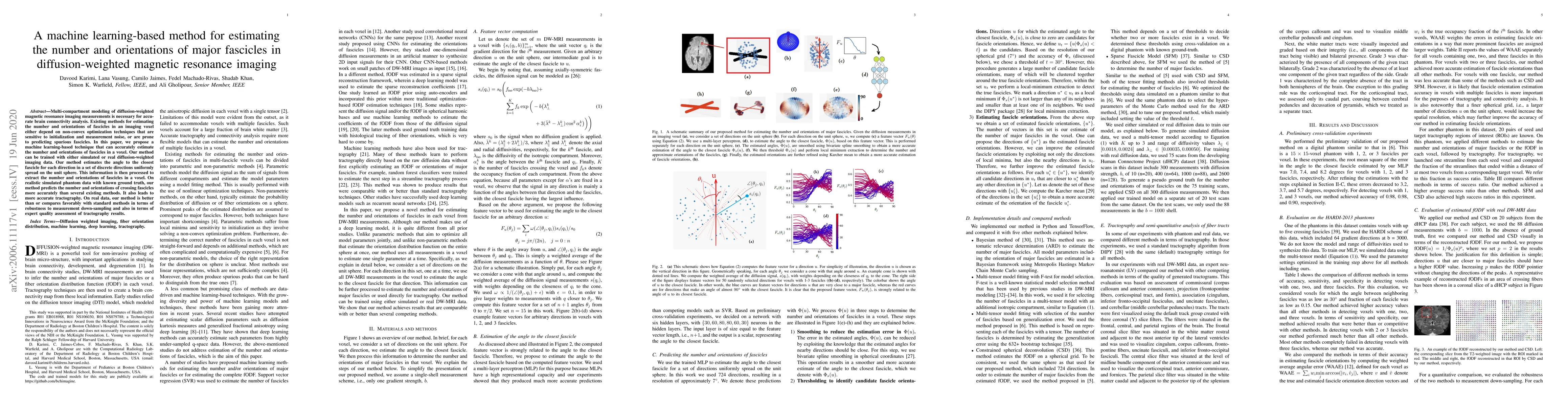
Multi-compartment modeling of diffusion-weighted magnetic resonance imaging measurements is necessary for accurate brain connectivity analysis. Existing methods for estimating the number and orienta...
Transfer learning is widely used for training machine learning models. Here, we study the role of transfer learning for training fully convolutional networks (FCNs) for medical image segmentation. O...
For single source helical Computed Tomography (CT), both Filtered-Back Projection (FBP) and statistical iterative reconstruction have been investigated. However for dual source CT with flying focal ...

Fast data acquisition in Magnetic Resonance Imaging (MRI) is vastly in demand and scan time directly depends on the number of acquired k-space samples. The data-driven methods based on deep neural n...
Fast data acquisition in Magnetic Resonance Imaging (MRI) is vastly in demand and scan time directly depends on the number of acquired k-space samples. Conventional MRI reconstruction methods for fa...
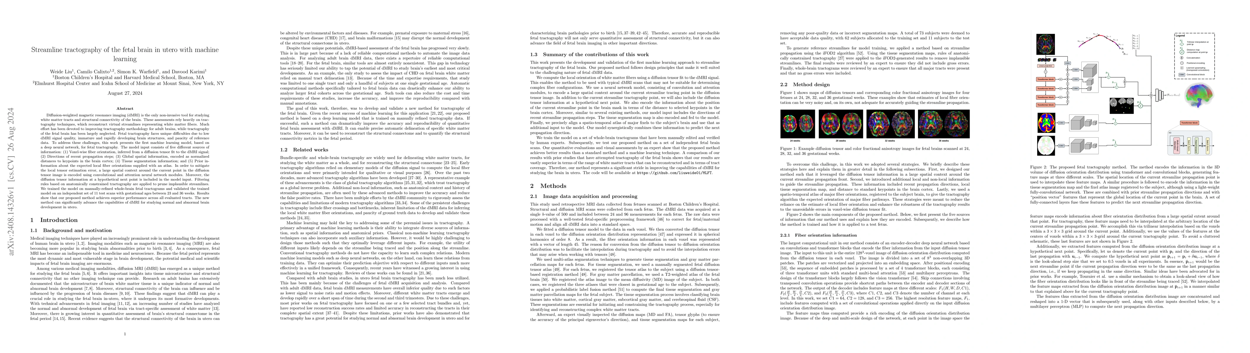
Diffusion-weighted magnetic resonance imaging (dMRI) is the only non-invasive tool for studying white matter tracts and structural connectivity of the brain. These assessments rely heavily on tractogr...
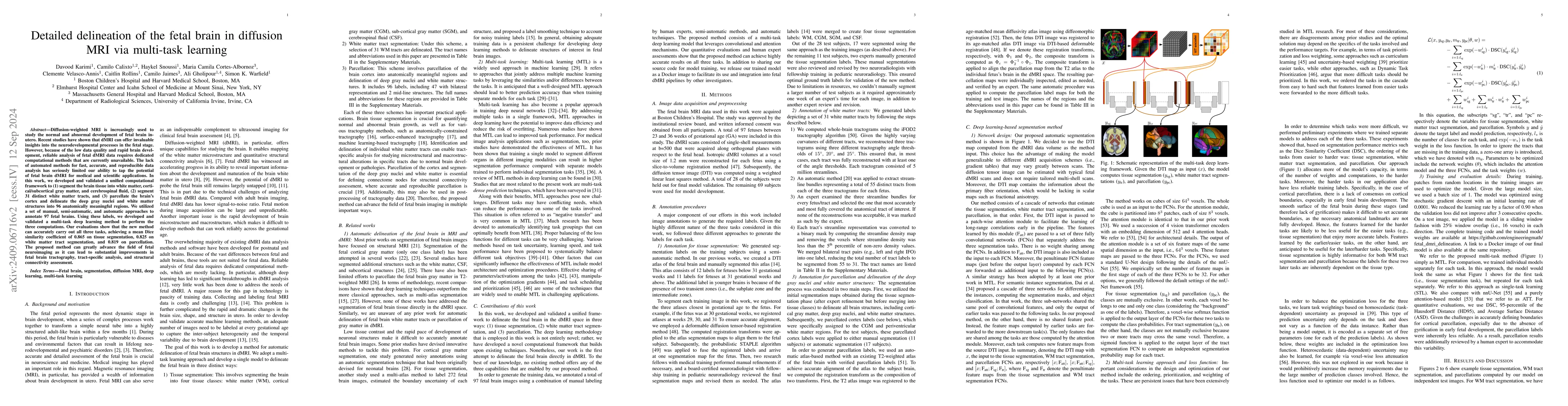
Diffusion-weighted MRI is increasingly used to study the normal and abnormal development of fetal brain in-utero. Recent studies have shown that dMRI can offer invaluable insights into the neurodevelo...
There is a growing interest in using diffusion MRI to study the white matter tracts and structural connectivity of the fetal brain. Recent progress in data acquisition and processing suggests that thi...
Purpose: To develop a rapid, high-resolution and distortion-free quantitative $R_{2}^{*}$ mapping technique for fetal brain at 3 T. Methods: A 2D multi-echo radial FLASH sequence with blip gradients...
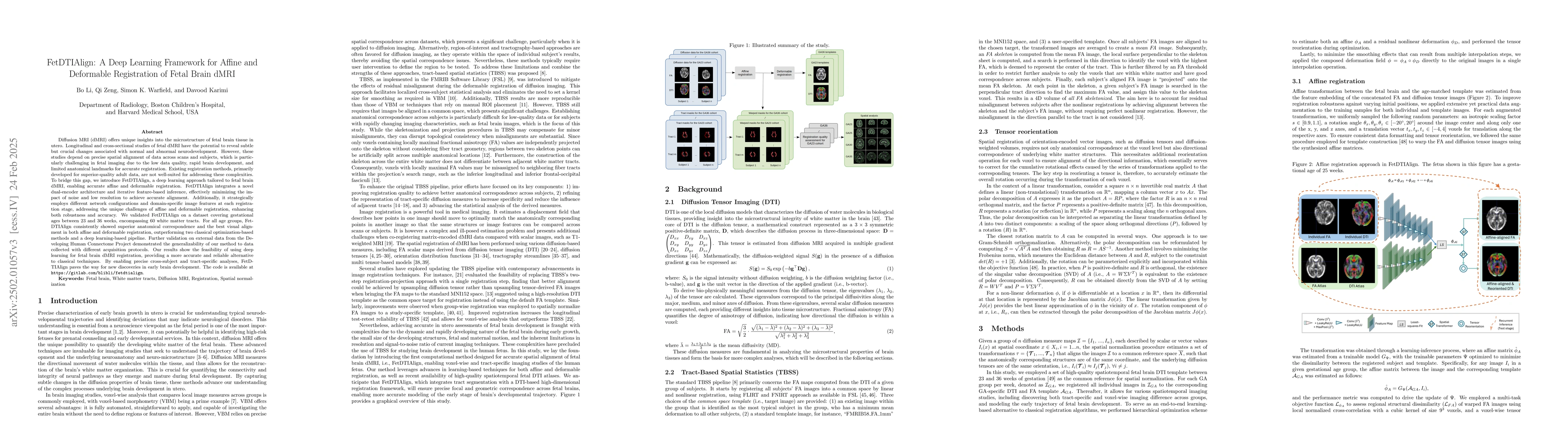
Diffusion MRI (dMRI) provides unique insights into fetal brain microstructure in utero. Longitudinal and cross-sectional fetal dMRI studies can reveal crucial neurodevelopmental changes but require pr...
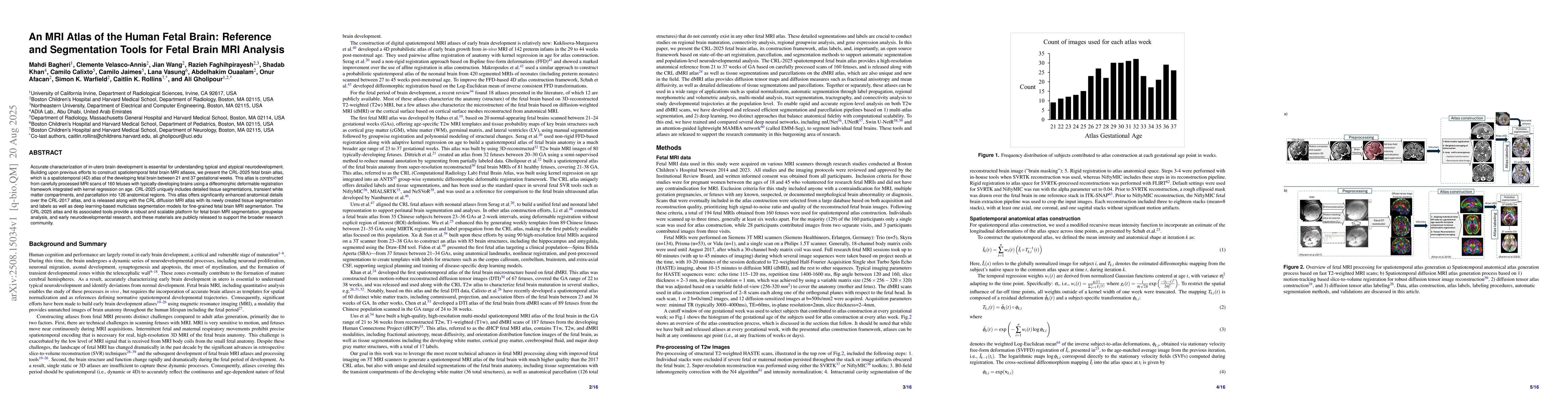
Accurate characterization of in-utero brain development is essential for understanding typical and atypical neurodevelopment. Building upon previous efforts to construct spatiotemporal fetal brain MRI...
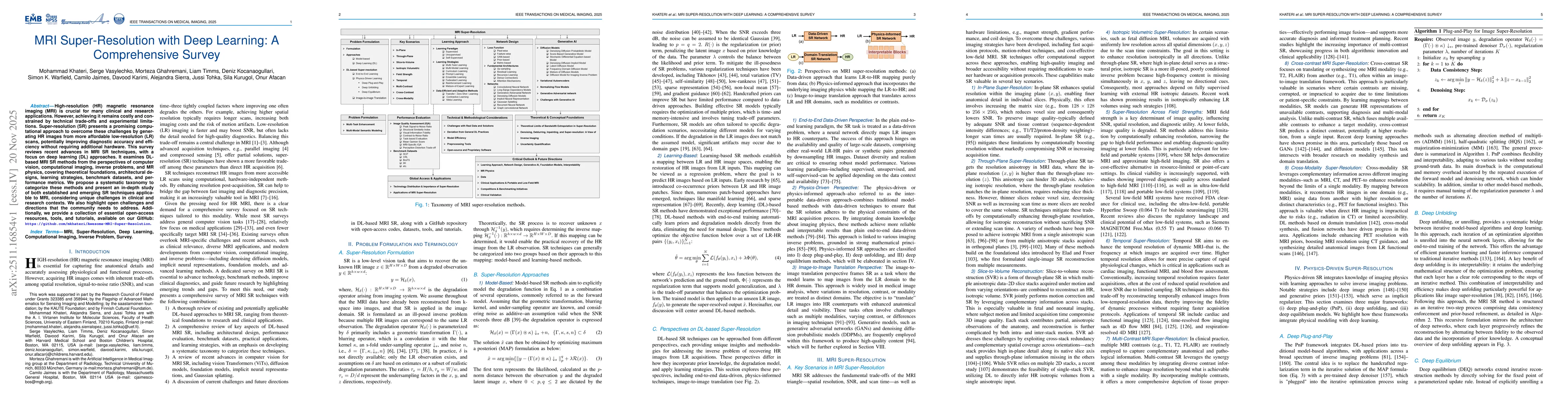
High-resolution (HR) magnetic resonance imaging (MRI) is crucial for many clinical and research applications. However, achieving it remains costly and constrained by technical trade-offs and experimen...
Background and Purpose: Automated detection of focal cortical dysplasia (FCD) requires large volumes of voxelwise lesion-delineated MRI data, which are difficult to acquire. This study aims to generat...