Stuart Crozier

13 papers on arXiv

Academic Profile

Name: Stuart Crozier
Total Papers: 1

Statistics

arXiv Papers 13
Total Publications 1

Papers on arXiv

Thumbnail for Automated anomaly-aware 3D segmentation of bones and cartilages in knee
  MR images from the Osteoarthritis Initiative

Automated anomaly-aware 3D segmentation of bones and cartilages in knee MR images from the Osteoarthritis Initiative

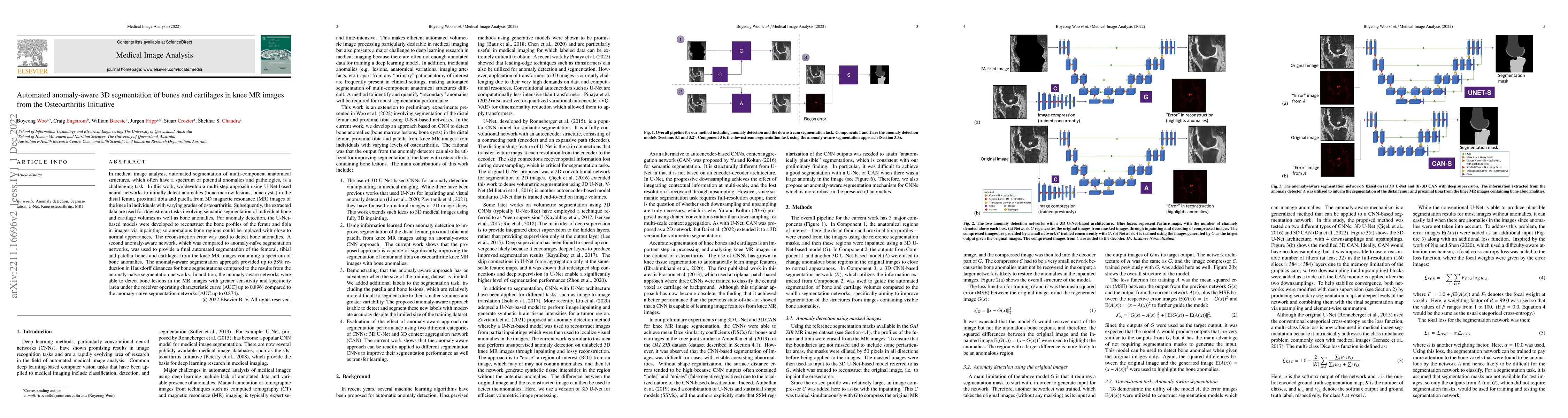
In medical image analysis, automated segmentation of multi-component anatomical structures, which often have a spectrum of potential anomalies and pathologies, is a challenging task. In this work, w...

Thumbnail for Structure Guided Manifolds for Discovery of Disease Characteristics

Structure Guided Manifolds for Discovery of Disease Characteristics

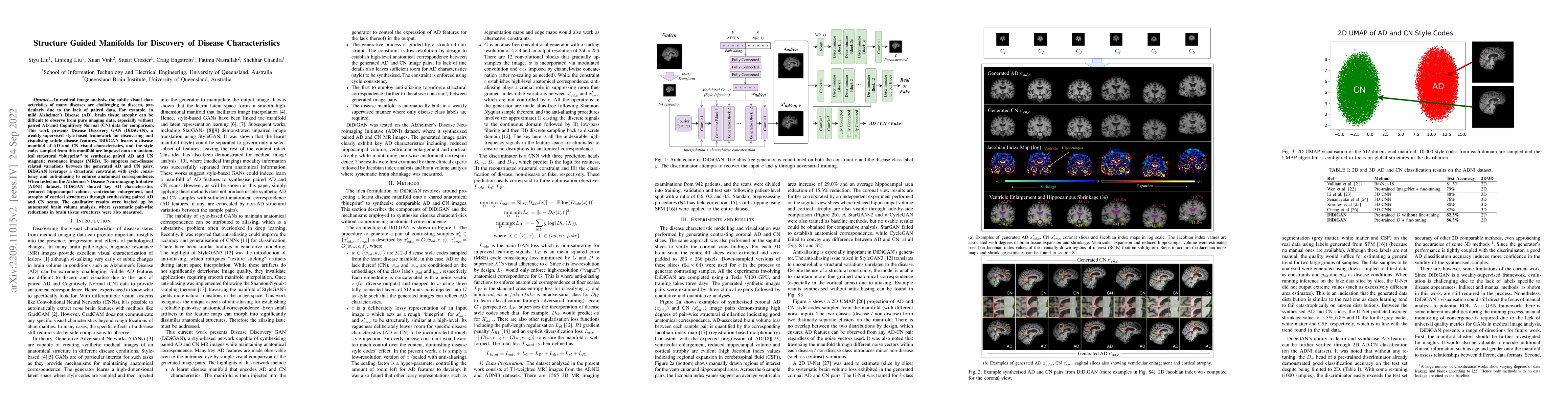
In medical image analysis, the subtle visual characteristics of many diseases are challenging to discern, particularly due to the lack of paired data. For example, in mild Alzheimer's Disease (AD), ...

Thumbnail for BFRnet: A deep learning-based MR background field removal method for QSM
  of the brain containing significant pathological susceptibility sources

BFRnet: A deep learning-based MR background field removal method for QSM of the brain containing significant pathological susceptibility sources

Introduction: Background field removal (BFR) is a critical step required for successful quantitative susceptibility mapping (QSM). However, eliminating the background field in brains containing sign...

Thumbnail for Automated volumetric and statistical shape assessment of cam-type
  morphology of the femoral head-neck region from 3D magnetic resonance images

Automated volumetric and statistical shape assessment of cam-type morphology of the femoral head-neck region from 3D magnetic resonance images

Femoroacetabular impingement (FAI) cam morphology is routinely assessed using two-dimensional alpha angles which do not provide specific data on cam size characteristics. The purpose of this study i...

Thumbnail for Instant tissue field and magnetic susceptibility mapping from MR raw
  phase using Laplacian enabled deep neural networks

Instant tissue field and magnetic susceptibility mapping from MR raw phase using Laplacian enabled deep neural networks

Quantitative susceptibility mapping (QSM) is a valuable MRI post-processing technique that quantifies the magnetic susceptibility of body tissue from phase data. However, the traditional QSM reconst...

Thumbnail for Bespoke Fractal Sampling Patterns for Discrete Fourier Space via the
  Kaleidoscope Transform

Bespoke Fractal Sampling Patterns for Discrete Fourier Space via the Kaleidoscope Transform

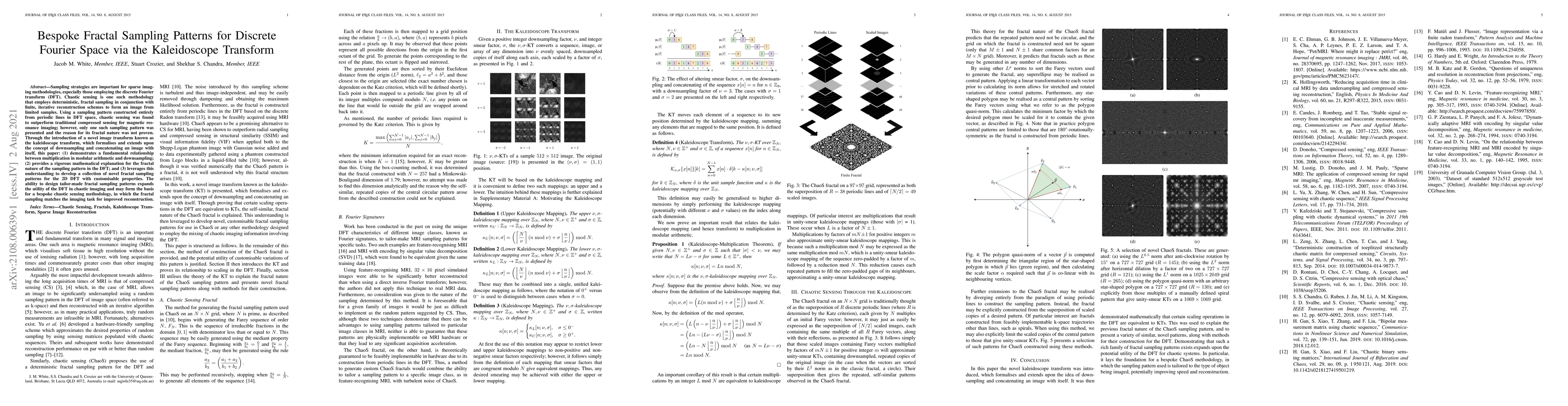
Sampling strategies are important for sparse imaging methodologies, especially those employing the discrete Fourier transform (DFT). Chaotic sensing is one such methodology that employs deterministi...

Thumbnail for Deep grey matter quantitative susceptibility mapping from small spatial
  coverages using deep learning

Deep grey matter quantitative susceptibility mapping from small spatial coverages using deep learning

Introduction: Quantitative Susceptibility Mapping (QSM) is generally acquired with full brain coverage, even though many QSM brain-iron studies focus on the deep grey matter (DGM) region only. Reduc...

Thumbnail for Deep Simultaneous Optimisation of Sampling and Reconstruction for
  Multi-contrast MRI

Deep Simultaneous Optimisation of Sampling and Reconstruction for Multi-contrast MRI

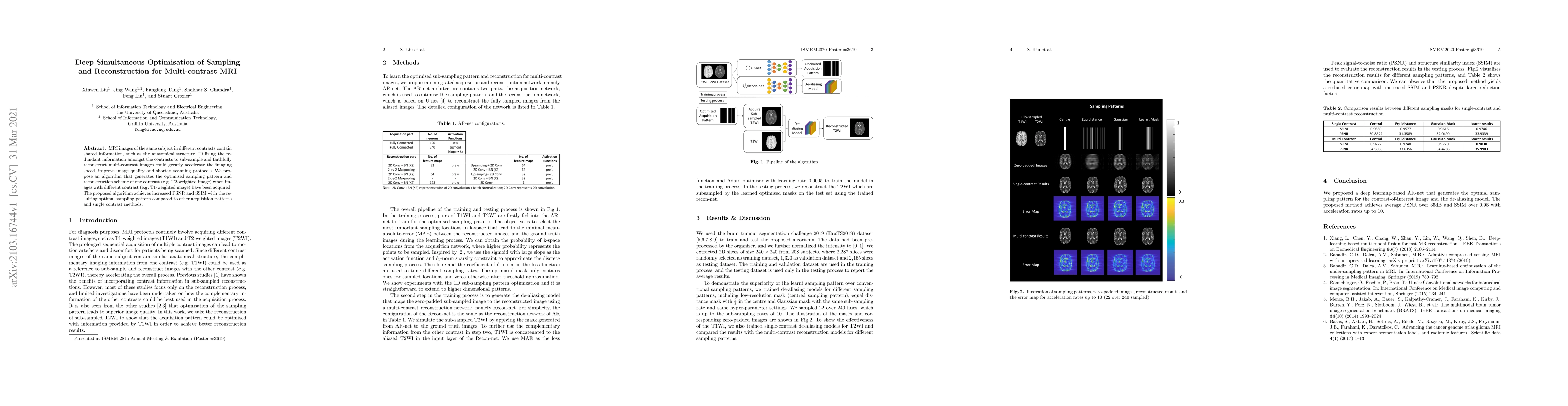
MRI images of the same subject in different contrasts contain shared information, such as the anatomical structure. Utilizing the redundant information amongst the contrasts to sub-sample and faithf...

Thumbnail for Accelerating Quantitative Susceptibility Mapping using Compressed
  Sensing and Deep Neural Network

Accelerating Quantitative Susceptibility Mapping using Compressed Sensing and Deep Neural Network

Quantitative susceptibility mapping (QSM) is an MRI phase-based post-processing method that quantifies tissue magnetic susceptibility distributions. However, QSM acquisitions are relatively slow, ev...

Thumbnail for Manipulating Medical Image Translation with Manifold Disentanglement

Manipulating Medical Image Translation with Manifold Disentanglement

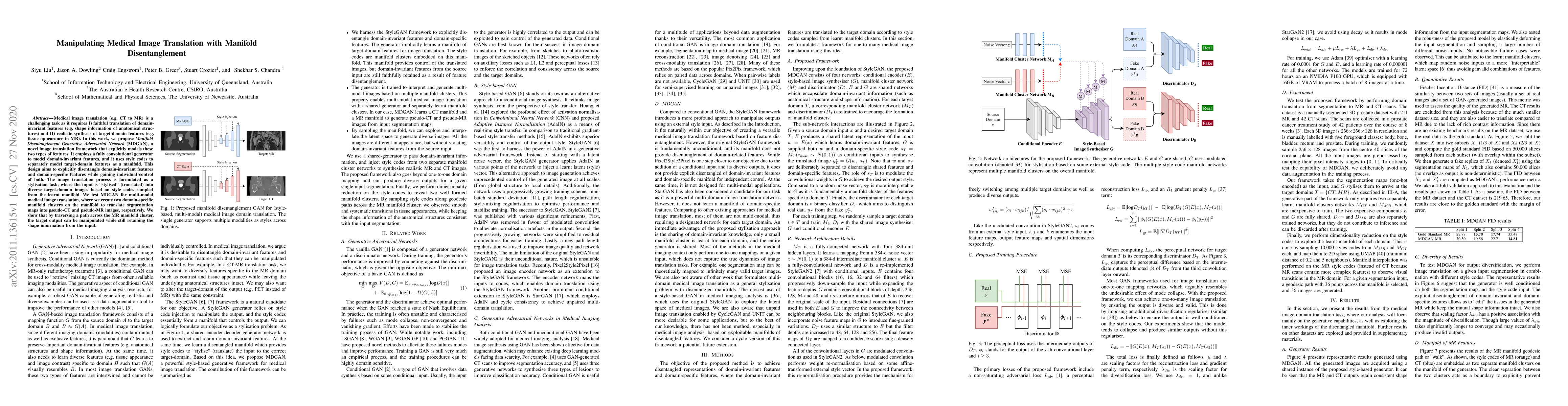
Medical image translation (e.g. CT to MR) is a challenging task as it requires I) faithful translation of domain-invariant features (e.g. shape information of anatomical structures) and II) realisti...

Thumbnail for Automatic lesion detection, segmentation and characterization via 3D
  multiscale morphological sifting in breast MRI

Automatic lesion detection, segmentation and characterization via 3D multiscale morphological sifting in breast MRI

Previous studies on computer aided detection/diagnosis (CAD) in 4D breast magnetic resonance imaging (MRI) regard lesion detection, segmentation and characterization as separate tasks, and typically...

Thumbnail for Generalisable 3D Fabric Architecture for Streamlined Universal
  Multi-Dataset Medical Image Segmentation

Generalisable 3D Fabric Architecture for Streamlined Universal Multi-Dataset Medical Image Segmentation

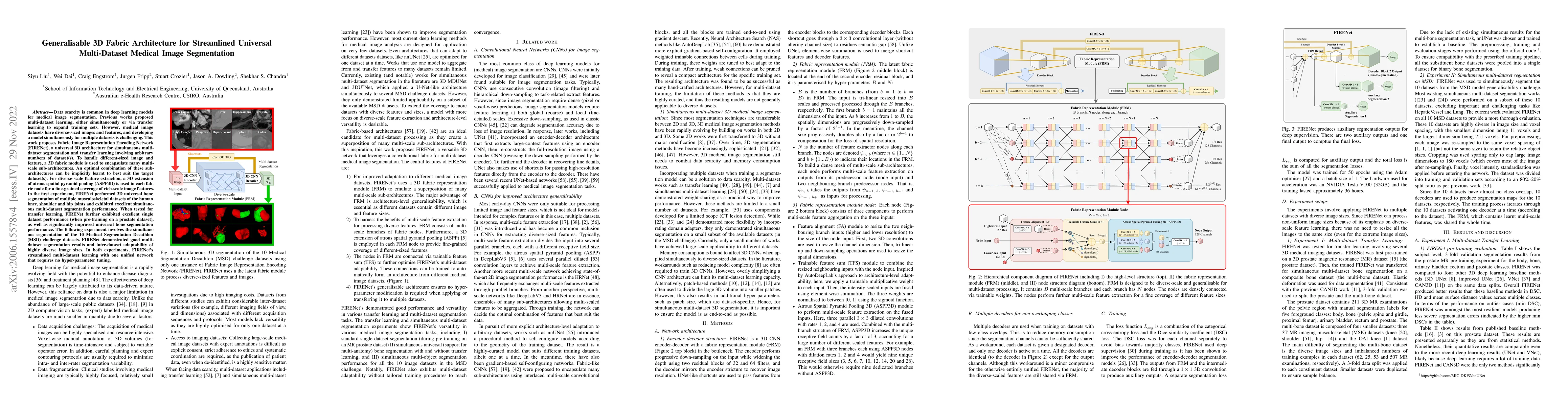
Data scarcity is common in deep learning models for medical image segmentation. Previous works proposed multi-dataset learning, either simultaneously or via transfer learning to expand training sets...

Thumbnail for Design and Construction of a Dedicated Radiolucent 8-element Flexible Radiofrequency (RF) Torso Coil for the 1.0T Australian MRI-Linac System

Design and Construction of a Dedicated Radiolucent 8-element Flexible Radiofrequency (RF) Torso Coil for the 1.0T Australian MRI-Linac System

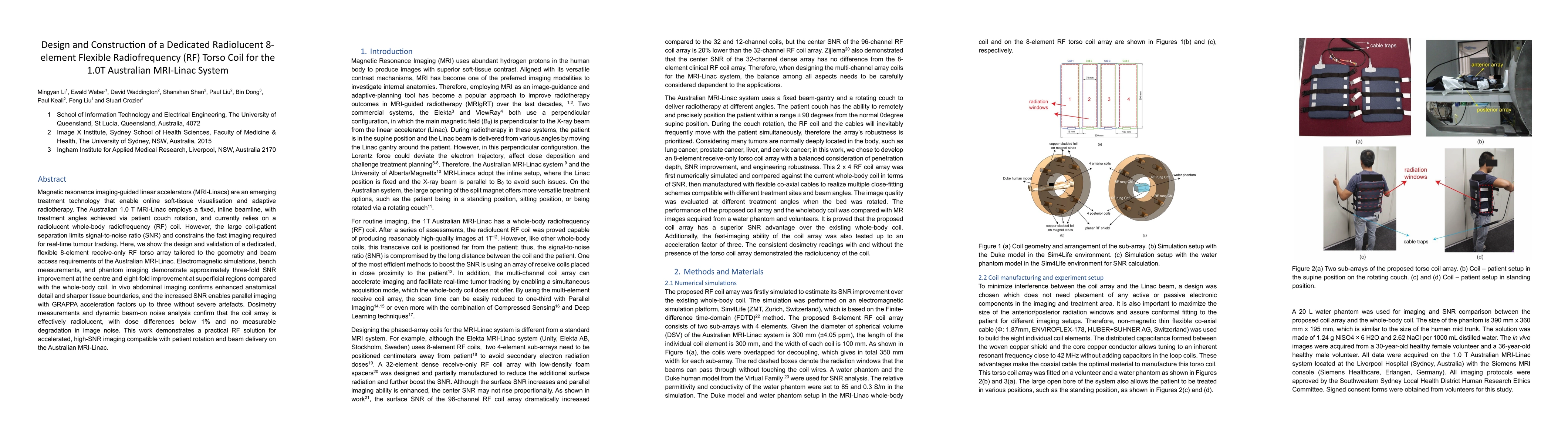
Magnetic resonance imaging-guided linear accelerators (MRI-Linacs) are an emerging treatment technology that enable online soft-tissue visualisation and adaptive radiotherapy. The Australian 1.0 T MRI...

This website uses cookies to ensure you get the best experience. Learn more