Theo van Walsum

13 papers on arXiv

Academic Profile

Name: T. Walsum
Total Papers: 165

Statistics

arXiv Papers 13
Total Publications 165

Papers on arXiv

Thumbnail for An automated framework for brain vessel centerline extraction from CTA
  images

An automated framework for brain vessel centerline extraction from CTA images

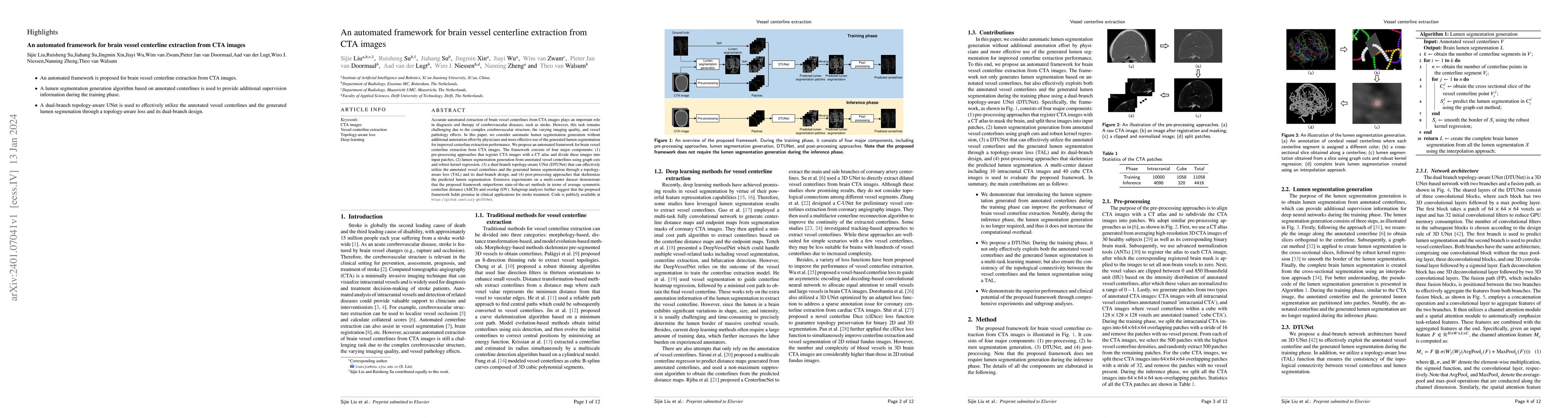
Accurate automated extraction of brain vessel centerlines from CTA images plays an important role in diagnosis and therapy of cerebrovascular diseases, such as stroke. However, this task remains cha...

Thumbnail for AngioMoCo: Learning-based Motion Correction in Cerebral Digital
  Subtraction Angiography

AngioMoCo: Learning-based Motion Correction in Cerebral Digital Subtraction Angiography

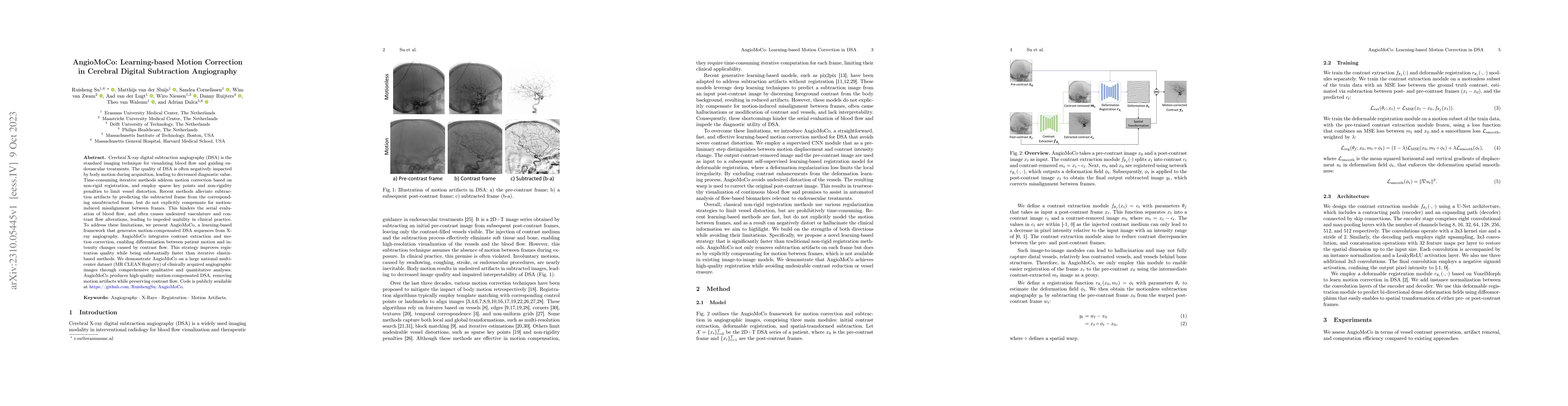
Cerebral X-ray digital subtraction angiography (DSA) is the standard imaging technique for visualizing blood flow and guiding endovascular treatments. The quality of DSA is often negatively impacted...

Thumbnail for Comparison of retinal regions-of-interest imaged by OCT for the
  classification of intermediate AMD

Comparison of retinal regions-of-interest imaged by OCT for the classification of intermediate AMD

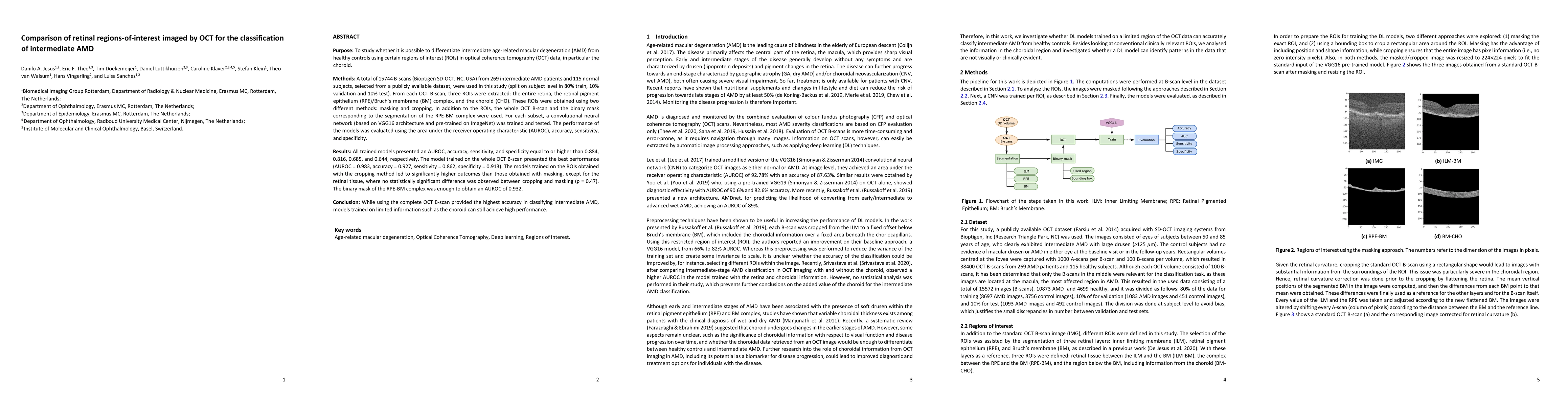
To study whether it is possible to differentiate intermediate age-related macular degeneration (AMD) from healthy controls using partial optical coherence tomography (OCT) data, that is, restricting...

Thumbnail for Label Refinement Network from Synthetic Error Augmentation for Medical
  Image Segmentation

Label Refinement Network from Synthetic Error Augmentation for Medical Image Segmentation

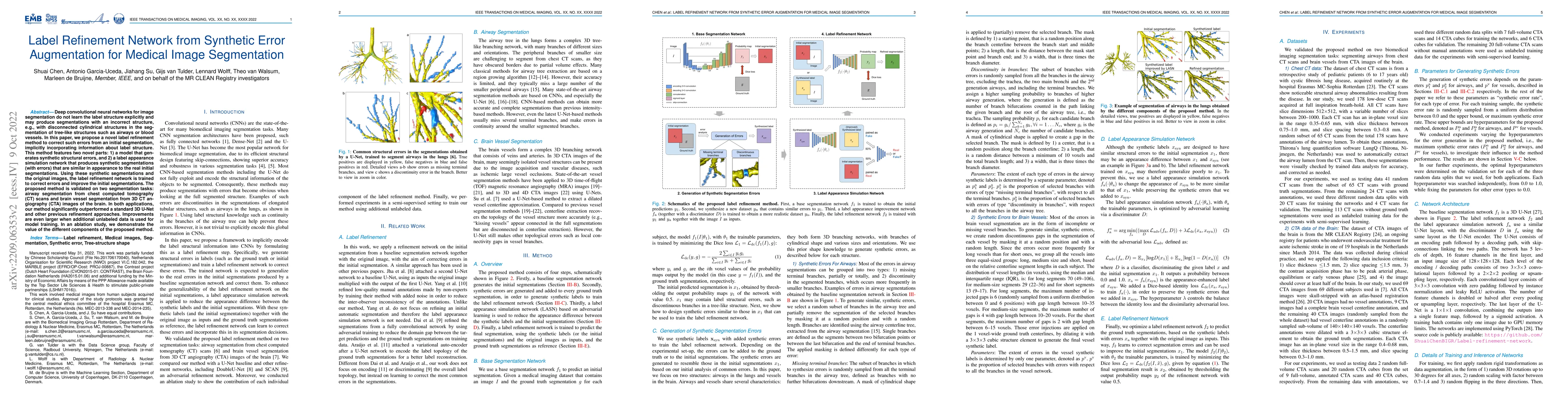
Deep convolutional neural networks for image segmentation do not learn the label structure explicitly and may produce segmentations with an incorrect structure, e.g., with disconnected cylindrical s...

Thumbnail for CAVE: Cerebral Artery-Vein Segmentation in Digital Subtraction
  Angiography

CAVE: Cerebral Artery-Vein Segmentation in Digital Subtraction Angiography

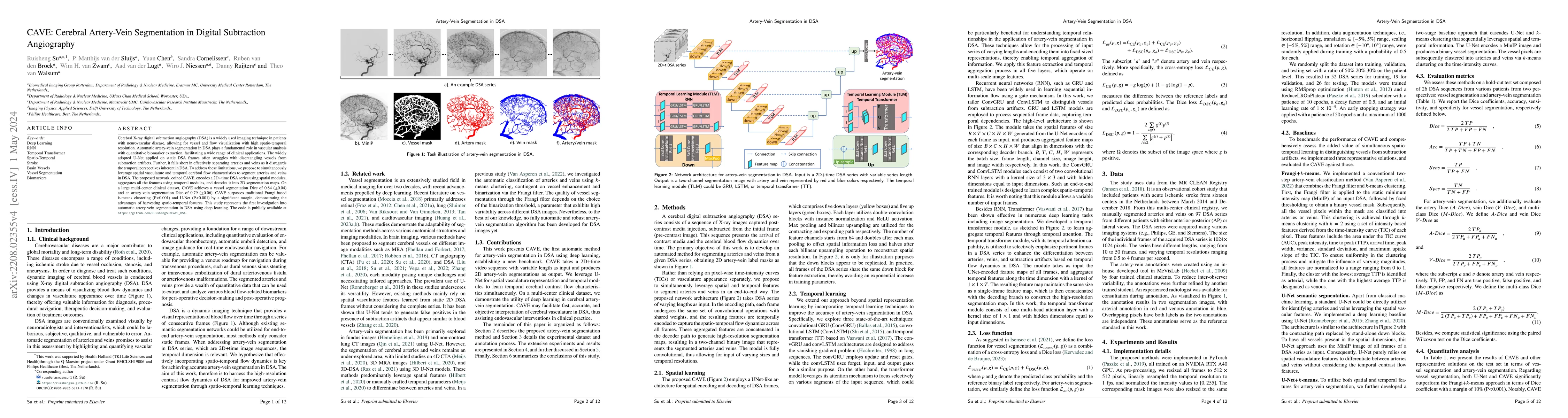
Cerebral X-ray digital subtraction angiography (DSA) is a widely used imaging technique in patients with neurovascular disease, allowing for vessel and flow visualization with high spatio-temporal r...

Thumbnail for Automatic Segmentation of the Optic Nerve Head Region in Optical
  Coherence Tomography: A Methodological Review

Automatic Segmentation of the Optic Nerve Head Region in Optical Coherence Tomography: A Methodological Review

The optic nerve head represents the intraocular section of the optic nerve (ONH), which is prone to damage by intraocular pressure. The advent of optical coherence tomography (OCT) has enabled the e...

Thumbnail for Signal-carrying speckle in Optical Coherence Tomography: a
  methodological review on biomedical applications

Signal-carrying speckle in Optical Coherence Tomography: a methodological review on biomedical applications

Significance: Speckle has historically been considered a source of noise in coherent light imaging. However, a number of works in optical coherence tomography (OCT) imaging have shown that speckle p...

Thumbnail for Dynamic Coronary Roadmapping via Catheter Tip Tracking in X-ray
  Fluoroscopy with Deep Learning Based Bayesian Filtering

Dynamic Coronary Roadmapping via Catheter Tip Tracking in X-ray Fluoroscopy with Deep Learning Based Bayesian Filtering

Percutaneous coronary intervention (PCI) is typically performed with image guidance using X-ray angiograms in which coronary arteries are opacified with X-ray opaque contrast agents. Interventional ...

Thumbnail for Fast Prospective Detection of Contrast Inflow in X-ray Angiograms with
  Convolutional Neural Network and Recurrent Neural Network

Fast Prospective Detection of Contrast Inflow in X-ray Angiograms with Convolutional Neural Network and Recurrent Neural Network

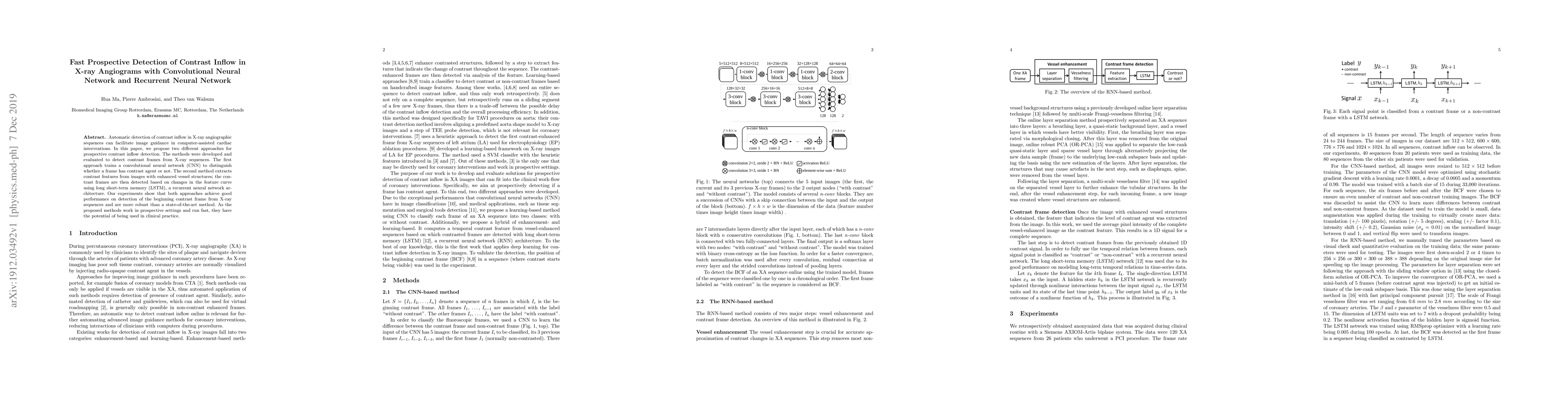
Automatic detection of contrast inflow in X-ray angiographic sequences can facilitate image guidance in computer-assisted cardiac interventions. In this paper, we propose two different approaches fo...

Thumbnail for Direct vascular territory segmentation on cerebral digital subtraction
  angiography

Direct vascular territory segmentation on cerebral digital subtraction angiography

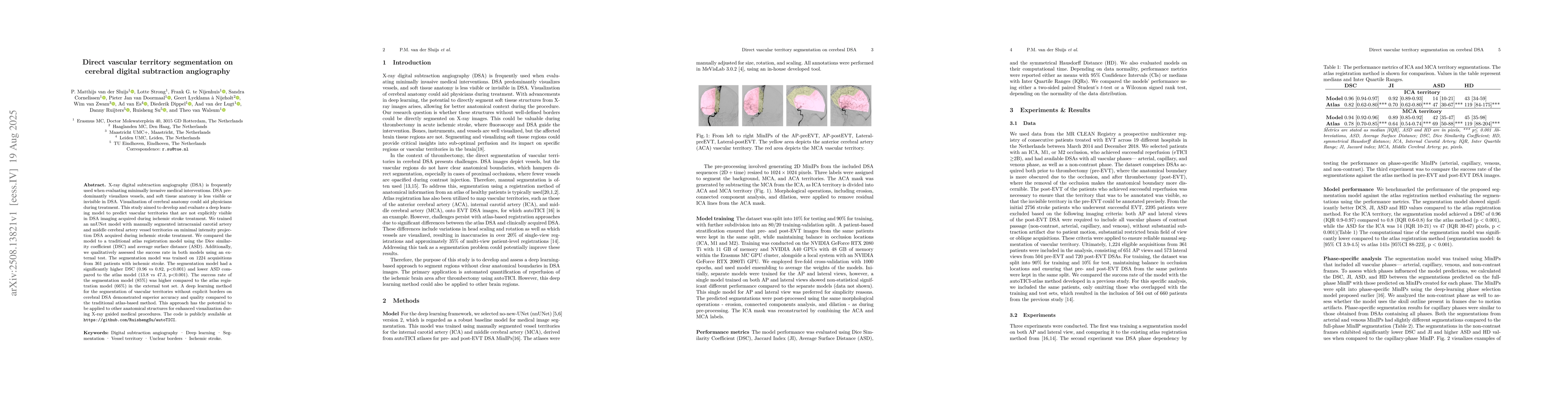
X-ray digital subtraction angiography (DSA) is frequently used when evaluating minimally invasive medical interventions. DSA predominantly visualizes vessels, and soft tissue anatomy is less visible o...

Thumbnail for OccluNet: Spatio-Temporal Deep Learning for Occlusion Detection on DSA

OccluNet: Spatio-Temporal Deep Learning for Occlusion Detection on DSA

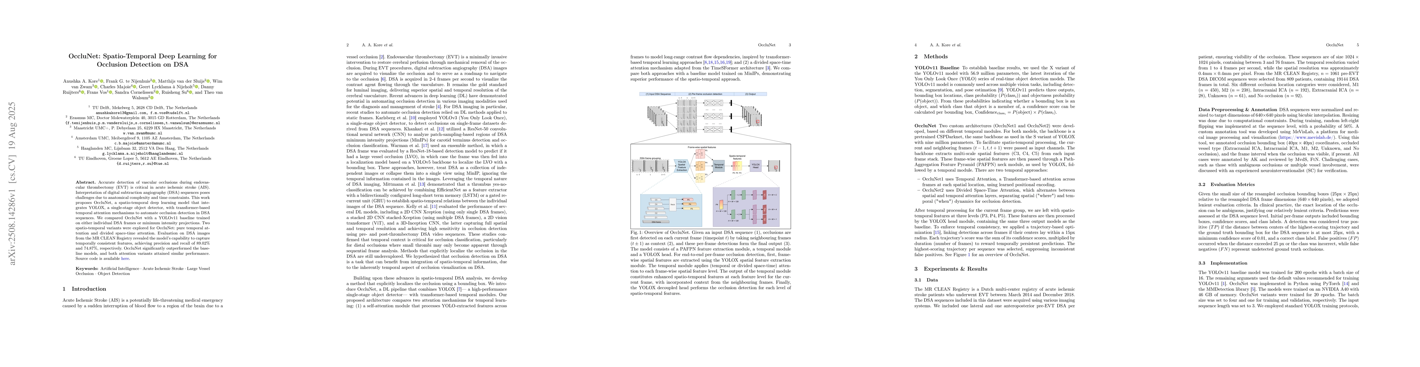
Accurate detection of vascular occlusions during endovascular thrombectomy (EVT) is critical in acute ischemic stroke (AIS). Interpretation of digital subtraction angiography (DSA) sequences poses cha...

Thumbnail for CLAIRE-DSA: Fluoroscopic Image Classification for Quality Assurance of
  Computer Vision Pipelines in Acute Ischemic Stroke

CLAIRE-DSA: Fluoroscopic Image Classification for Quality Assurance of Computer Vision Pipelines in Acute Ischemic Stroke

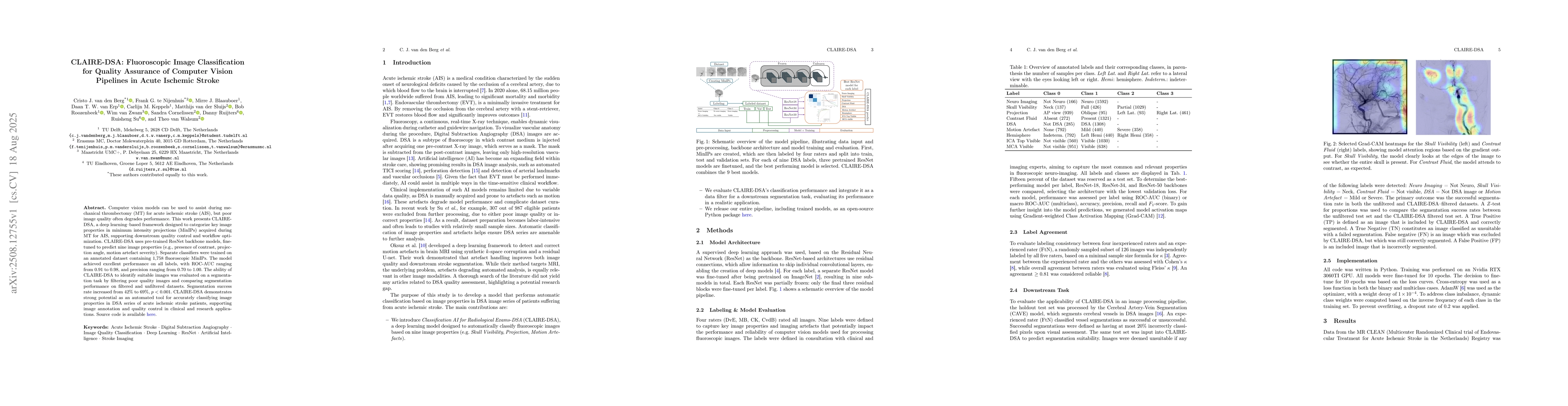
Computer vision models can be used to assist during mechanical thrombectomy (MT) for acute ischemic stroke (AIS), but poor image quality often degrades performance. This work presents CLAIRE-DSA, a de...

Thumbnail for SurgNavAR: An Augmented Reality Surgical Navigation Framework for Optical See-Through Head Mounted Displays

SurgNavAR: An Augmented Reality Surgical Navigation Framework for Optical See-Through Head Mounted Displays

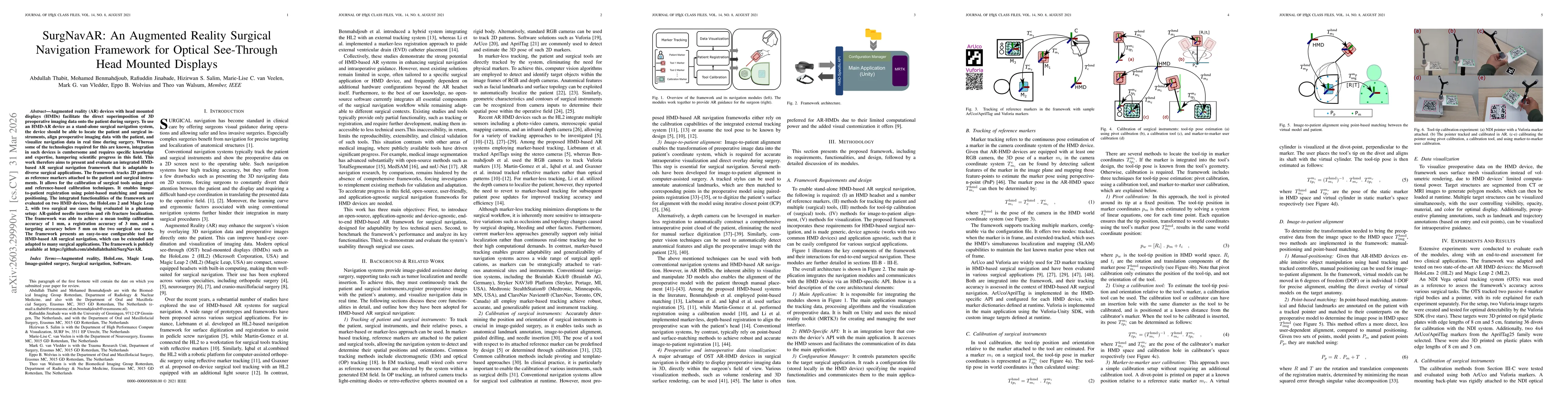
Augmented reality (AR) devices with head mounted displays (HMDs) facilitate the direct superimposition of 3D preoperative imaging data onto the patient during surgery. To use an HMD-AR device as a sta...

This website uses cookies to ensure you get the best experience. Learn more