Academic Profile
Statistics
Similar Authors
Papers on arXiv
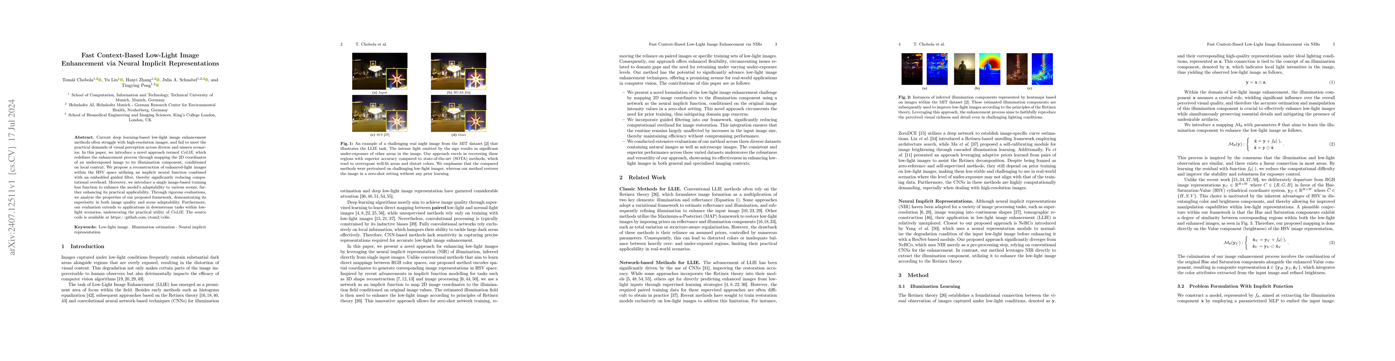
Current deep learning-based low-light image enhancement methods often struggle with high-resolution images, and fail to meet the practical demands of visual perception across diverse and unseen scenar...
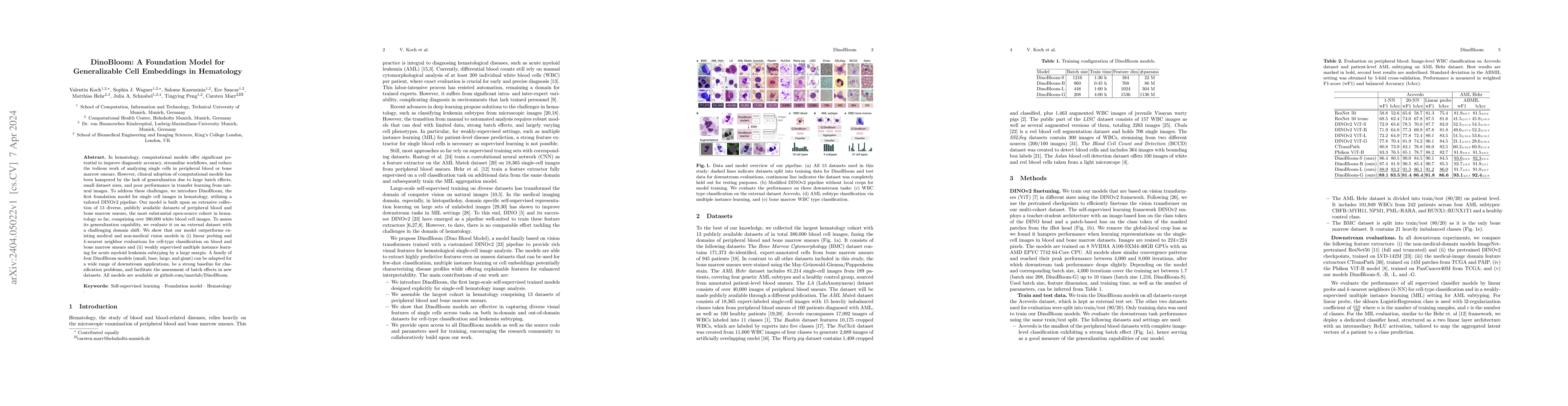
In hematology, computational models offer significant potential to improve diagnostic accuracy, streamline workflows, and reduce the tedious work of analyzing single cells in peripheral blood or bon...
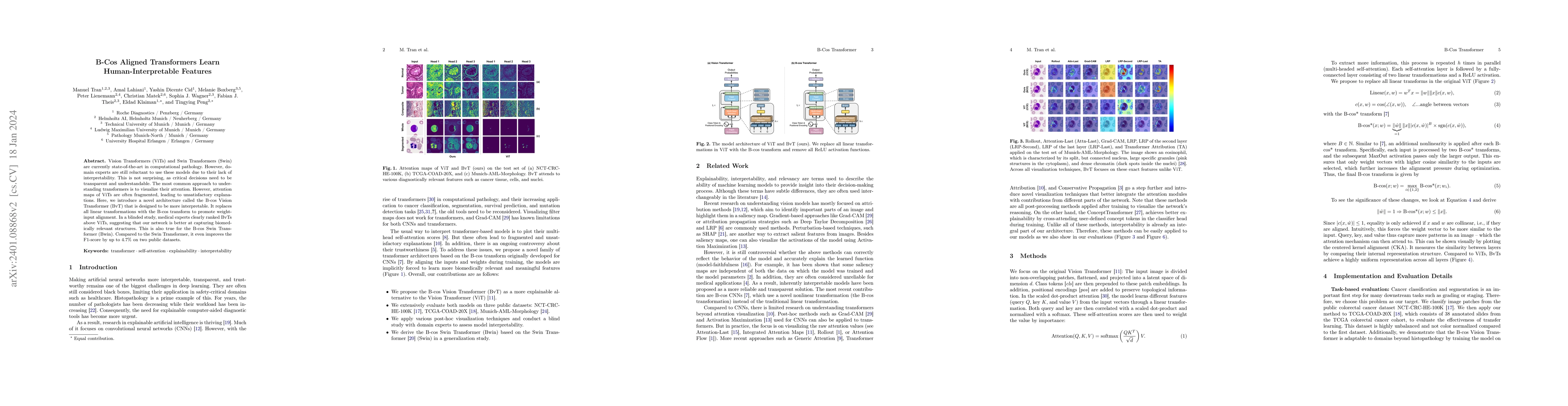
Vision Transformers (ViTs) and Swin Transformers (Swin) are currently state-of-the-art in computational pathology. However, domain experts are still reluctant to use these models due to their lack o...
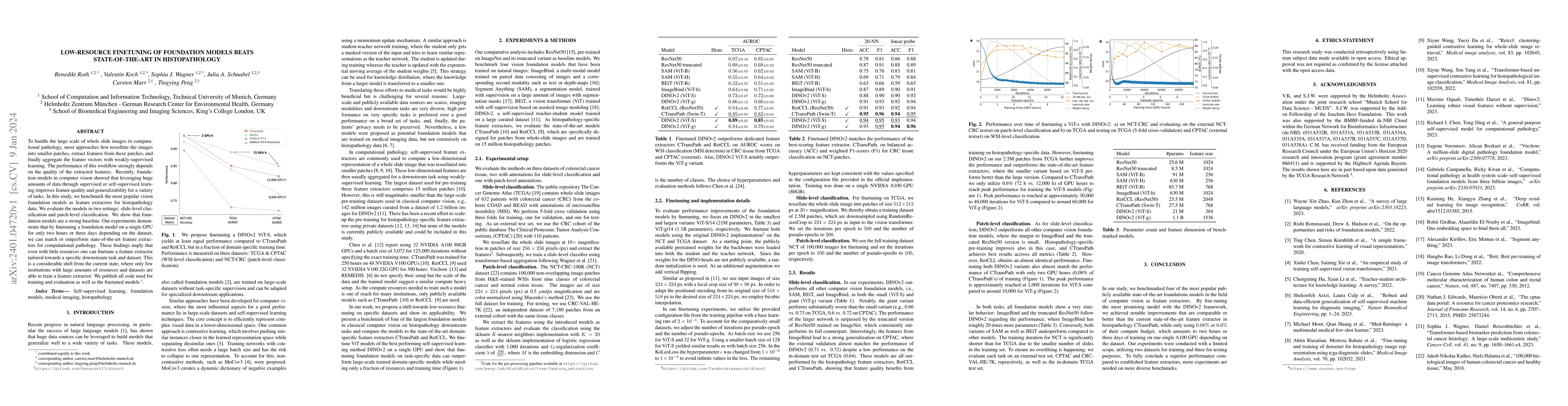
To handle the large scale of whole slide images in computational pathology, most approaches first tessellate the images into smaller patches, extract features from these patches, and finally aggrega...
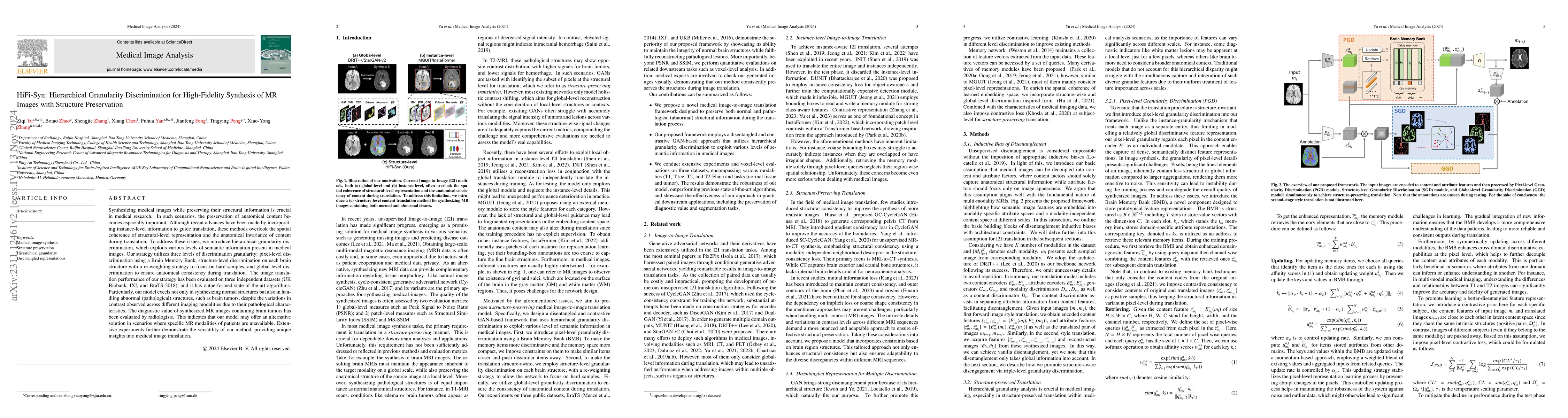
Synthesizing medical images while preserving their structural information is crucial in medical research. In such scenarios, the preservation of anatomical content becomes especially important. Alth...
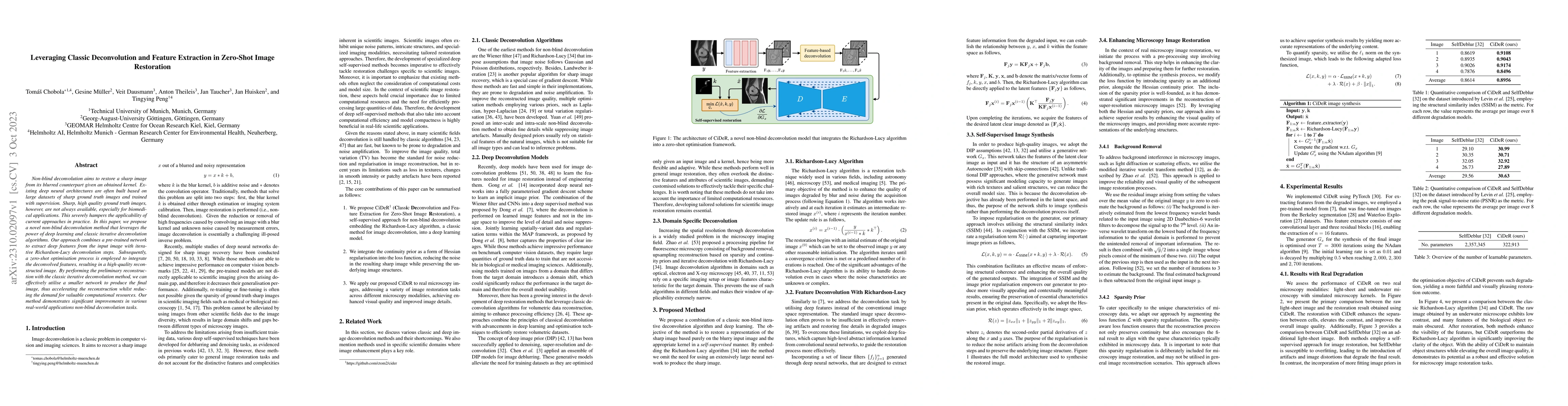
Non-blind deconvolution aims to restore a sharp image from its blurred counterpart given an obtained kernel. Existing deep neural architectures are often built based on large datasets of sharp groun...
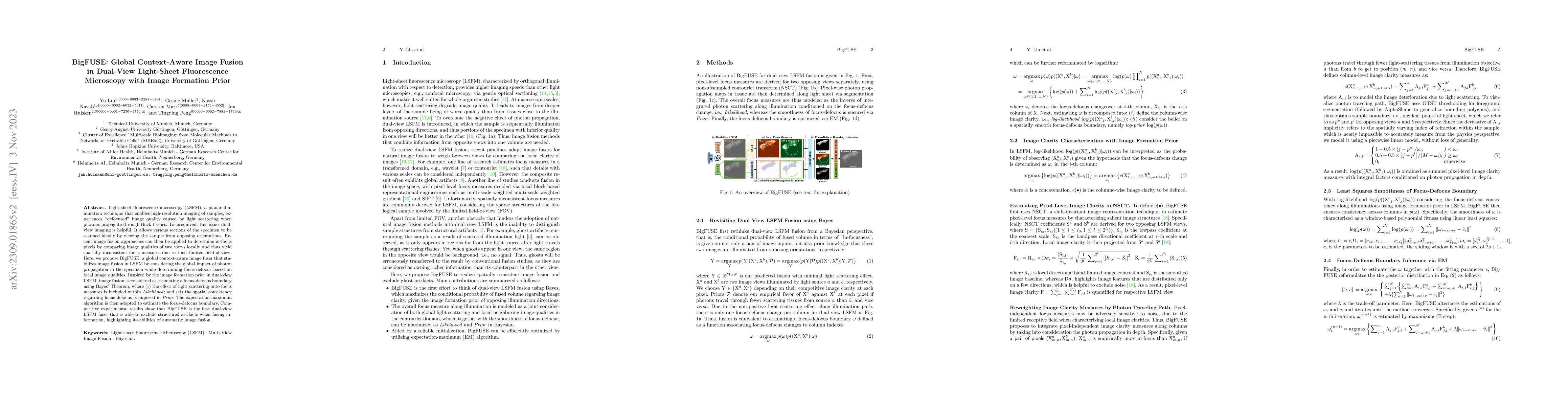
Light-sheet fluorescence microscopy (LSFM), a planar illumination technique that enables high-resolution imaging of samples, experiences defocused image quality caused by light scattering when photo...
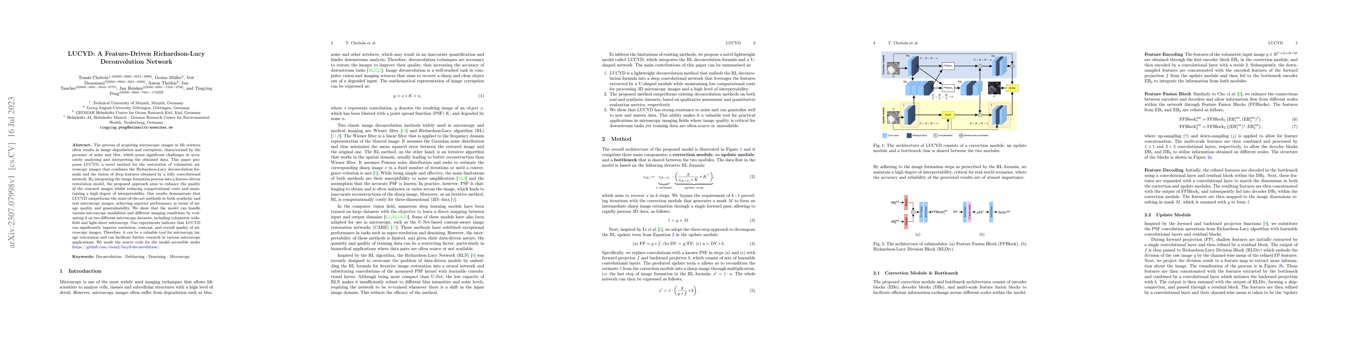
The process of acquiring microscopic images in life sciences often results in image degradation and corruption, characterised by the presence of noise and blur, which poses significant challenges in...
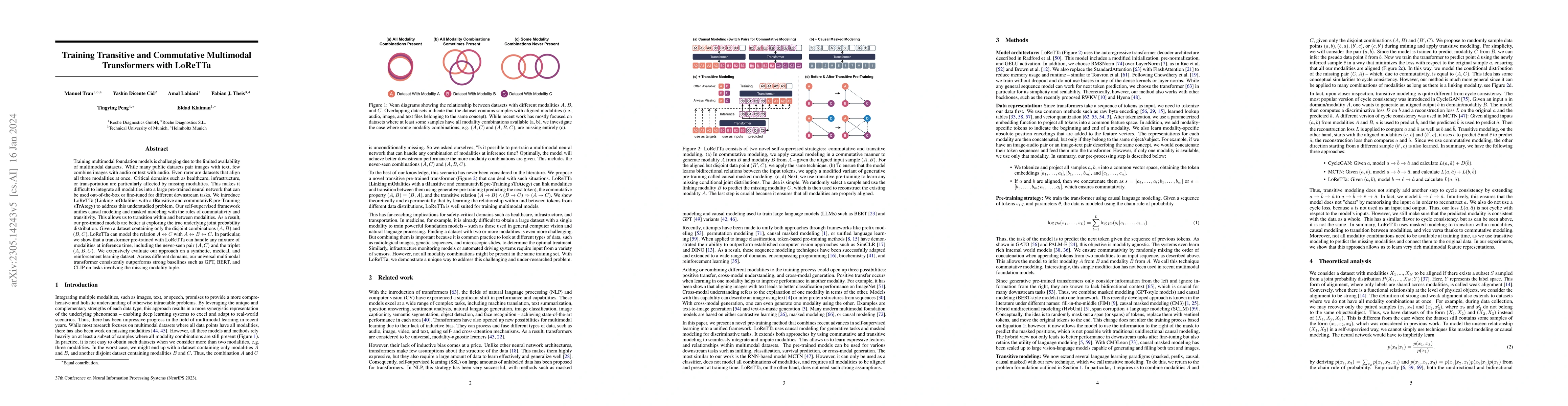
Training multimodal foundation models is challenging due to the limited availability of multimodal datasets. While many public datasets pair images with text, few combine images with audio or text w...
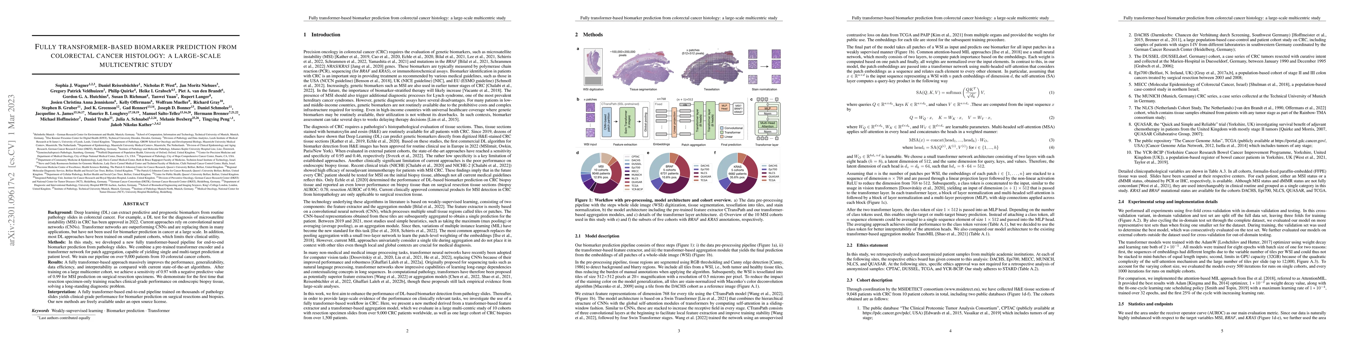
Background: Deep learning (DL) can extract predictive and prognostic biomarkers from routine pathology slides in colorectal cancer. For example, a DL test for the diagnosis of microsatellite instabi...
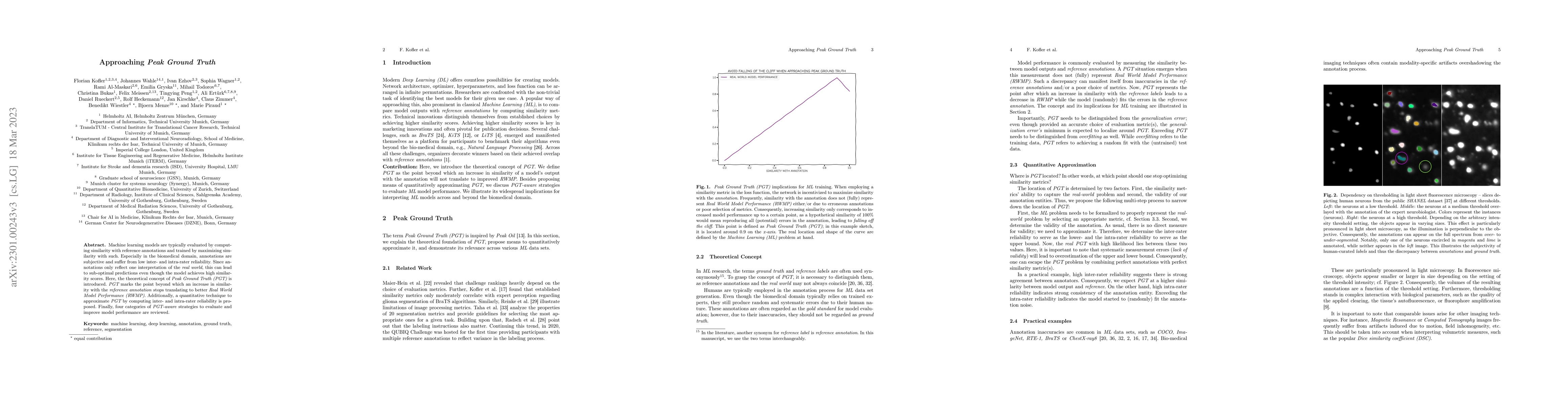
Machine learning models are typically evaluated by computing similarity with reference annotations and trained by maximizing similarity with such. Especially in the biomedical domain, annotations ar...
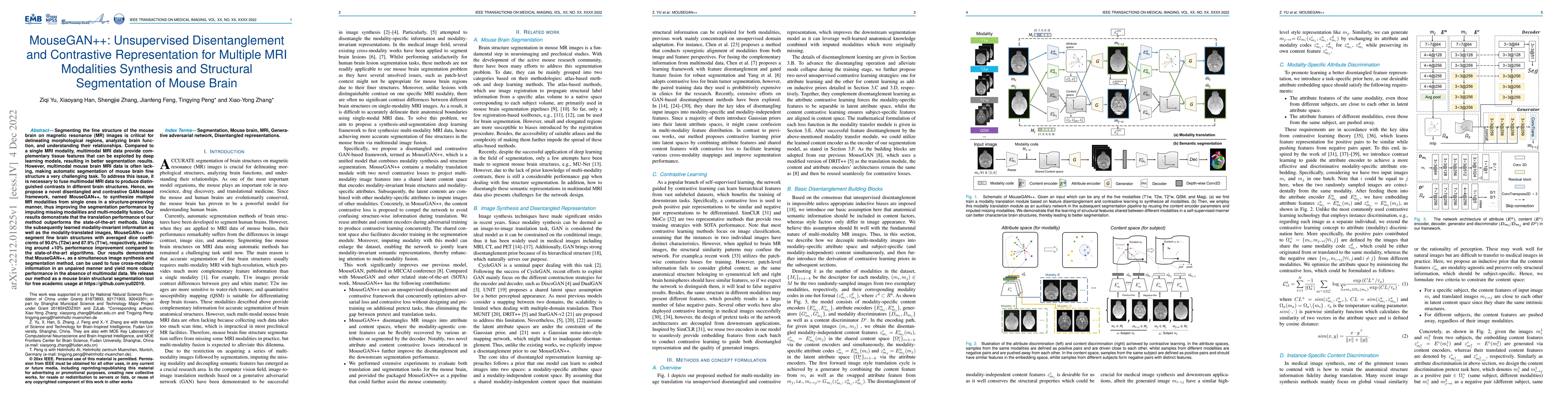
Segmenting the fine structure of the mouse brain on magnetic resonance (MR) images is critical for delineating morphological regions, analyzing brain function, and understanding their relationships....
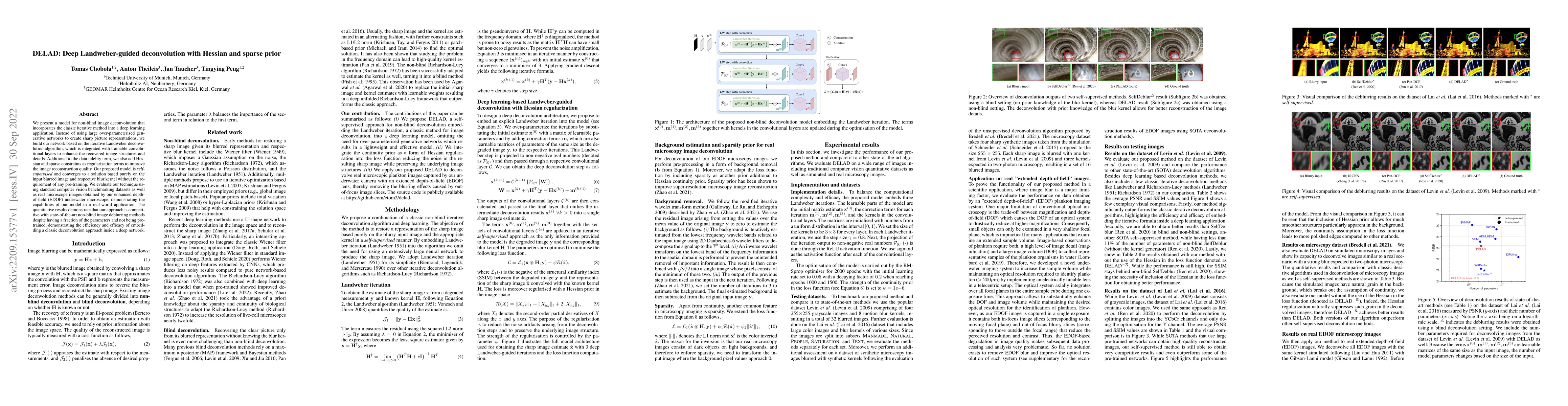
We present a model for non-blind image deconvolution that incorporates the classic iterative method into a deep learning application. Instead of using large over-parameterised generative networks to...
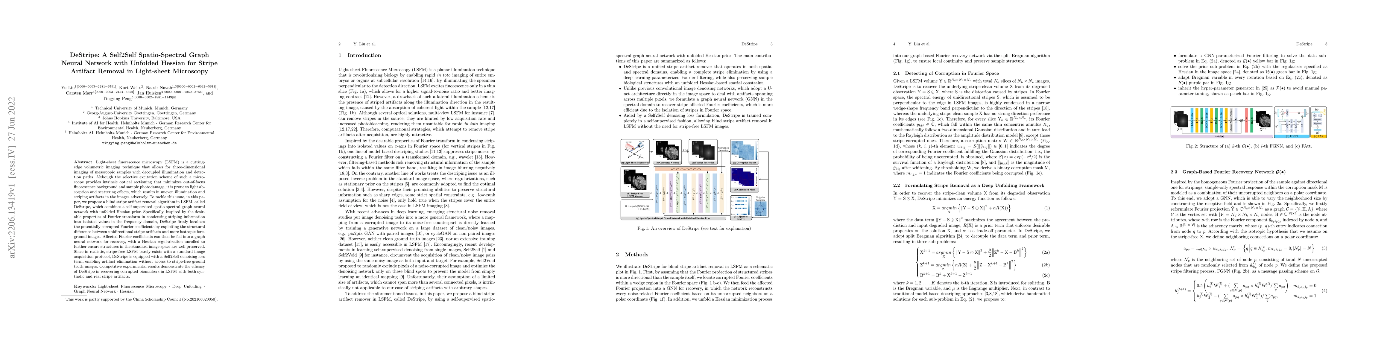
Light-sheet fluorescence microscopy (LSFM) is a cutting-edge volumetric imaging technique that allows for three-dimensional imaging of mesoscopic samples with decoupled illumination and detection pa...
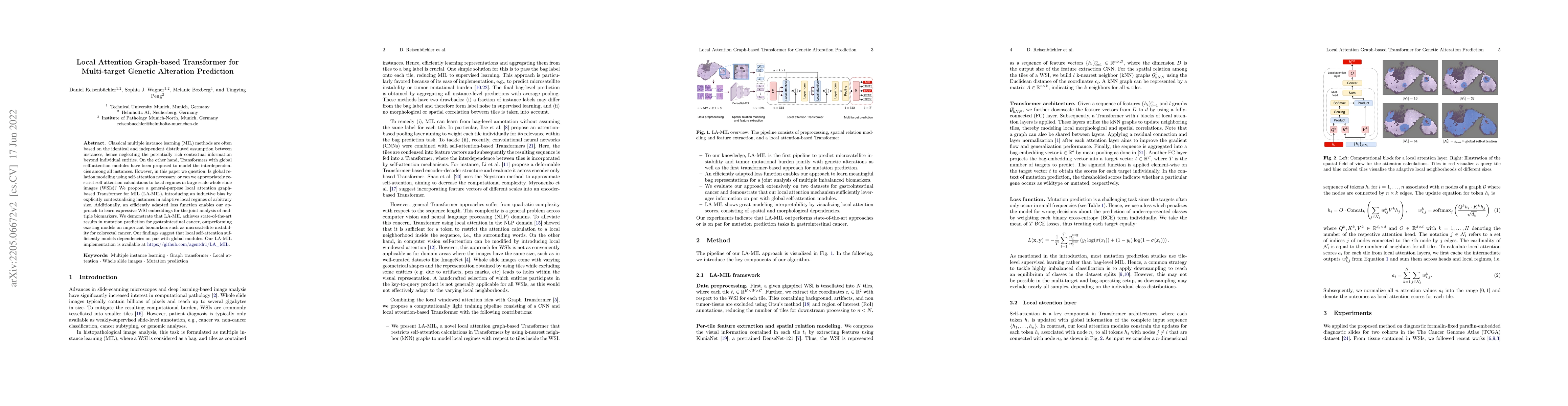
Classical multiple instance learning (MIL) methods are often based on the identical and independent distributed assumption between instances, hence neglecting the potentially rich contextual informa...
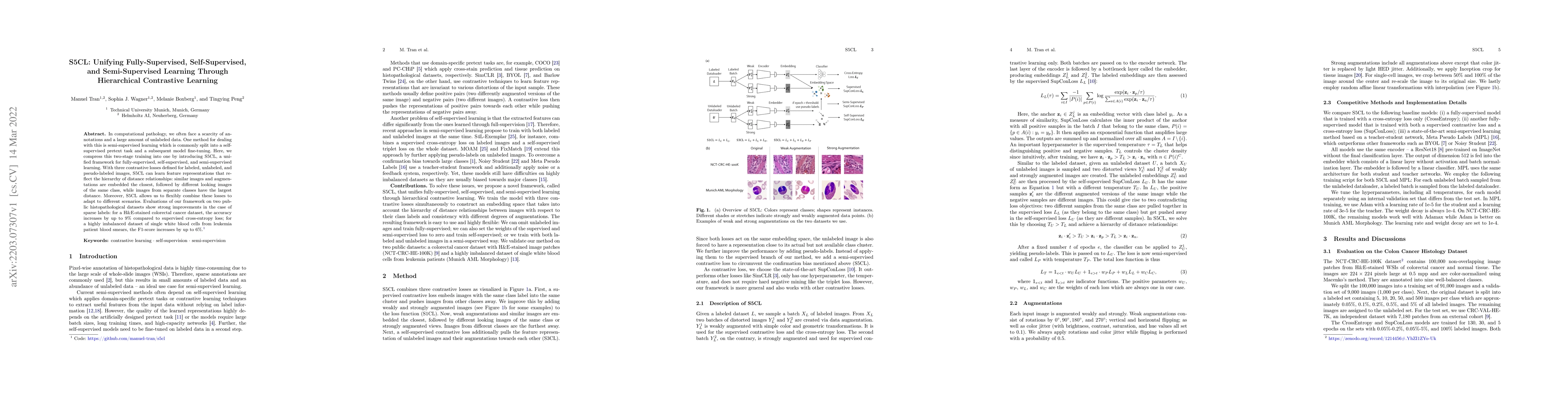
In computational pathology, we often face a scarcity of annotations and a large amount of unlabeled data. One method for dealing with this is semi-supervised learning which is commonly split into a ...
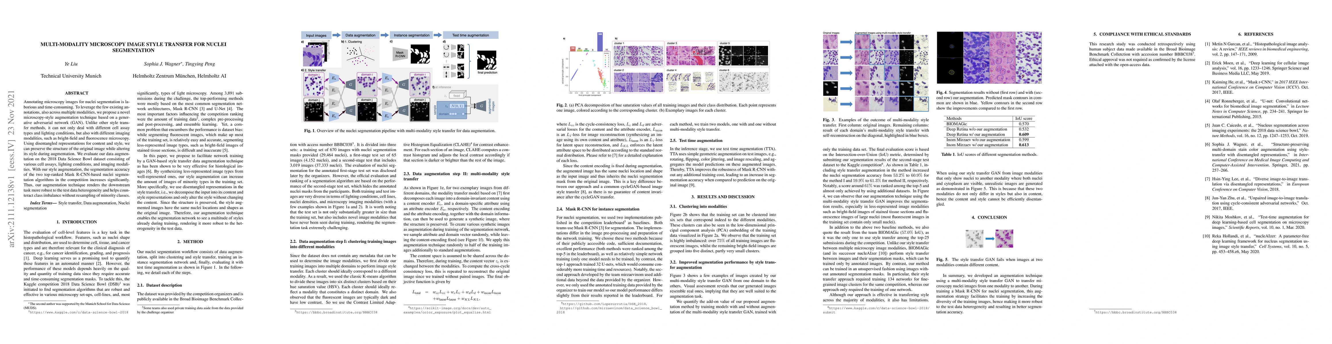
Annotating microscopy images for nuclei segmentation is laborious and time-consuming. To leverage the few existing annotations, also across multiple modalities, we propose a novel microscopy-style a...
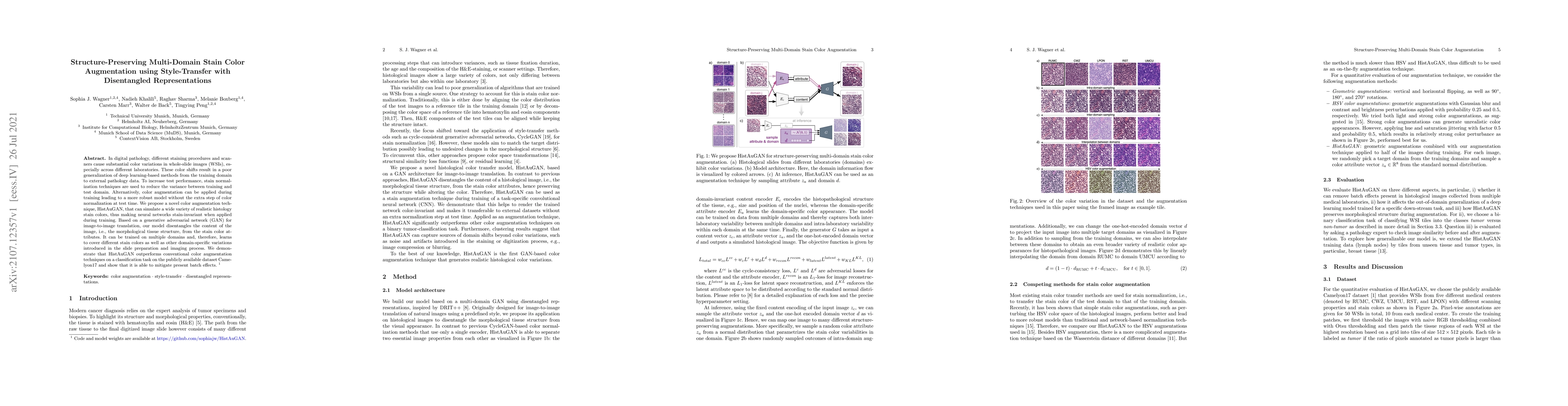
In digital pathology, different staining procedures and scanners cause substantial color variations in whole-slide images (WSIs), especially across different laboratories. These color shifts result ...
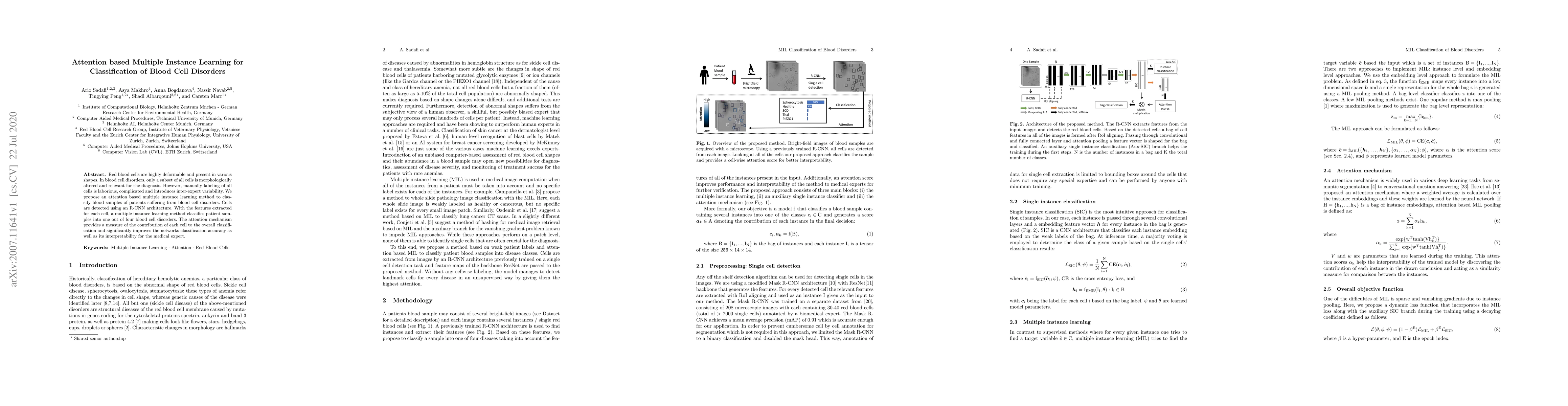
Red blood cells are highly deformable and present in various shapes. In blood cell disorders, only a subset of all cells is morphologically altered and relevant for the diagnosis. However, manually ...
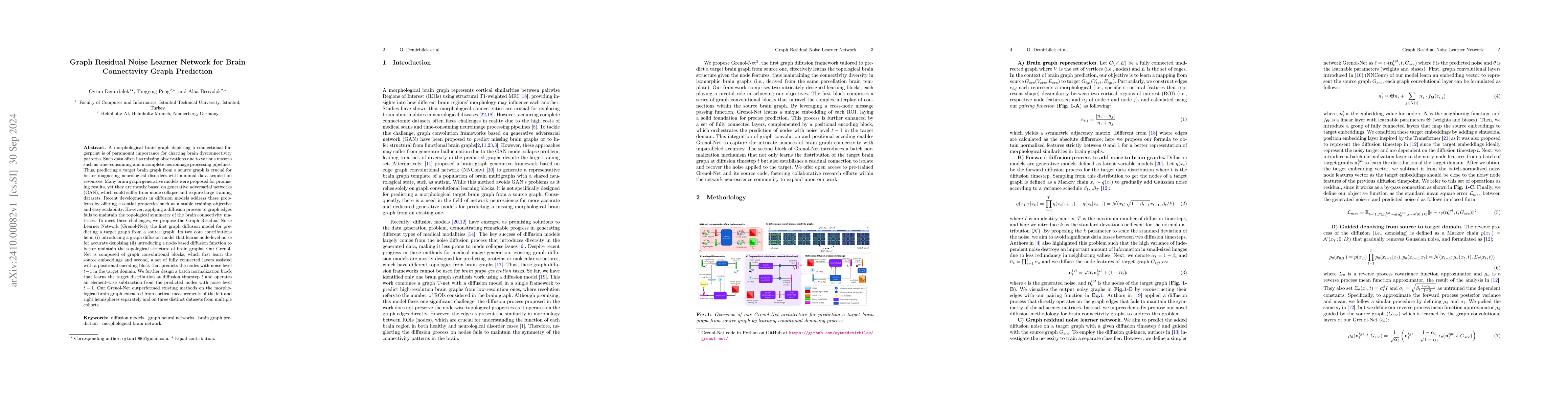
A morphological brain graph depicting a connectional fingerprint is of paramount importance for charting brain dysconnectivity patterns. Such data often has missing observations due to various reasons...
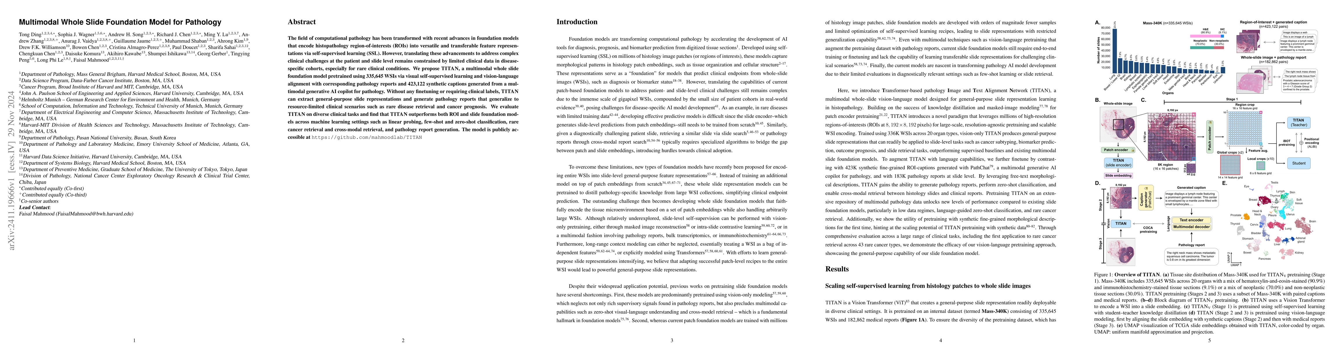
The field of computational pathology has been transformed with recent advances in foundation models that encode histopathology region-of-interests (ROIs) into versatile and transferable feature repres...
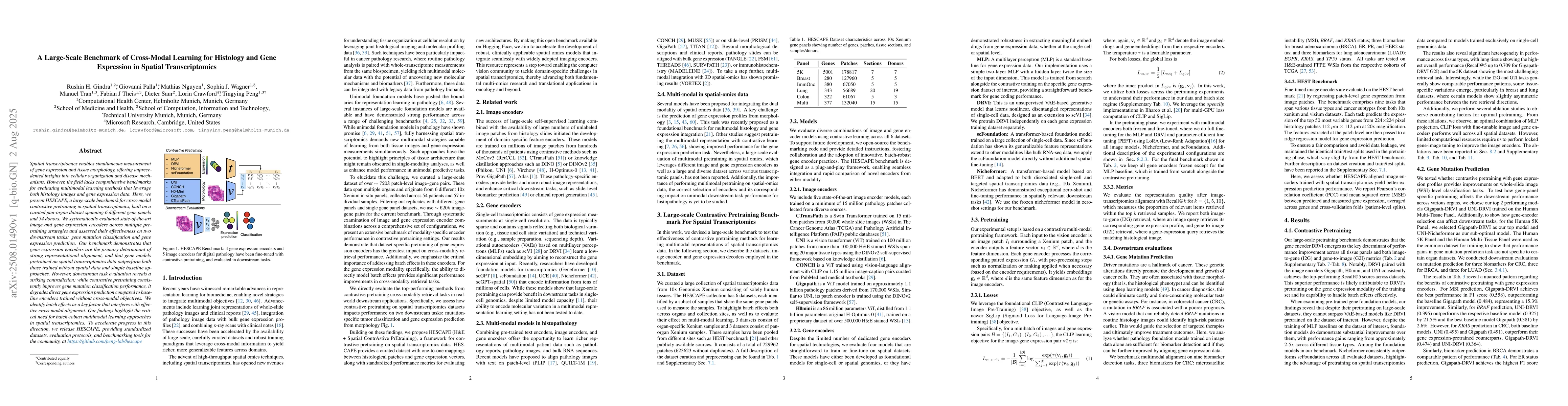
Spatial transcriptomics enables simultaneous measurement of gene expression and tissue morphology, offering unprecedented insights into cellular organization and disease mechanisms. However, the field...
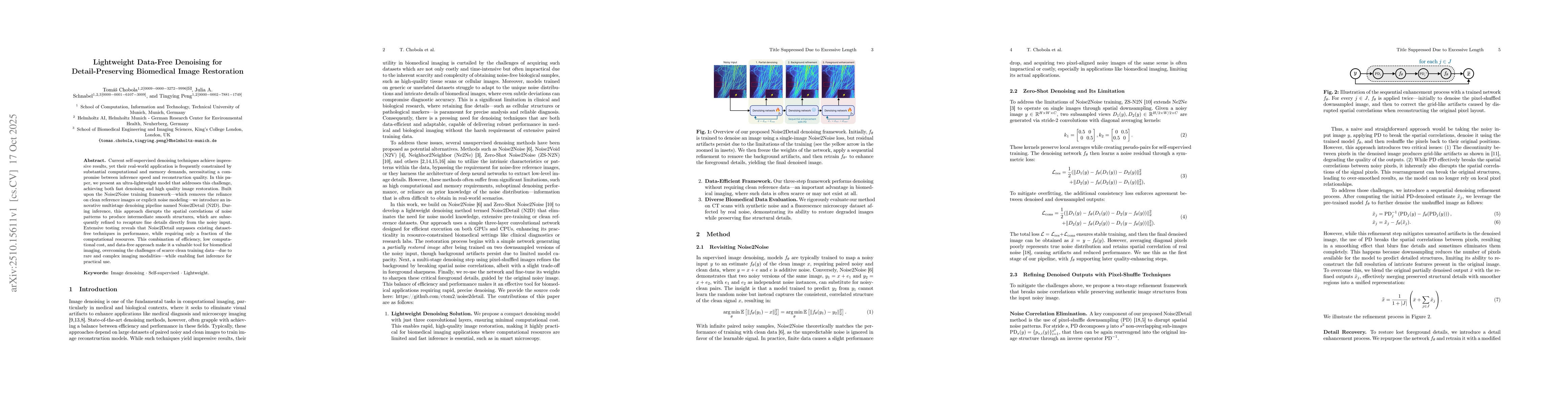
Current self-supervised denoising techniques achieve impressive results, yet their real-world application is frequently constrained by substantial computational and memory demands, necessitating a com...
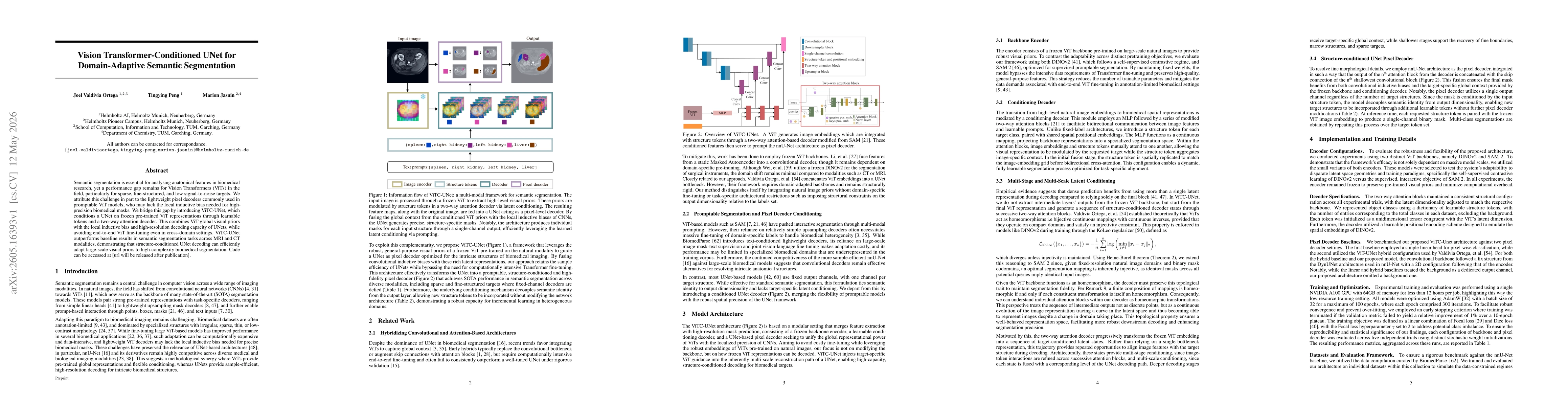
Semantic segmentation is essential for analysing anatomical features in biomedical research, yet a performance gap remains for Vision Transformers (ViTs) in the field, particularly for sparse, fine-st...
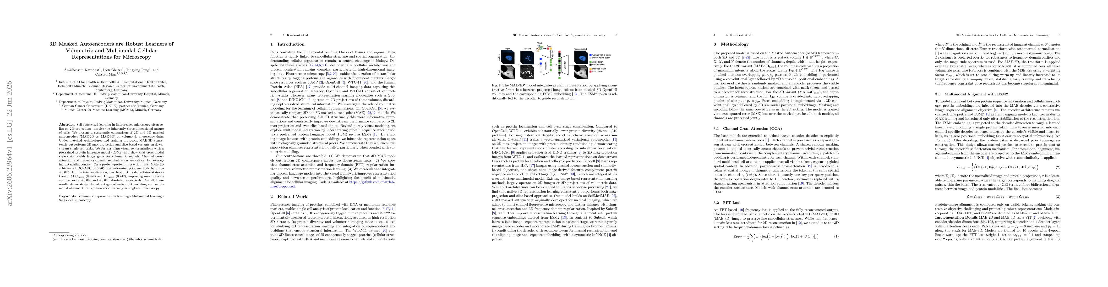
Self-supervised learning in fluorescence microscopy often relies on 2D projections, despite the inherently three-dimensional nature of cells. We present a systematic comparison of 2D and 3D masked aut...