Toru Nagasaka

4 papers on arXiv

Academic Profile

Name: T. Nagasaka
Total Papers: 5

Statistics

arXiv Papers 4
Total Publications 5

Papers on arXiv

Thumbnail for Novel Methods for Analyzing Cellular Interactions in Deep Learning-Based
  Image Cytometry: Spatial Interaction Potential and Co-Localization Index

Novel Methods for Analyzing Cellular Interactions in Deep Learning-Based Image Cytometry: Spatial Interaction Potential and Co-Localization Index

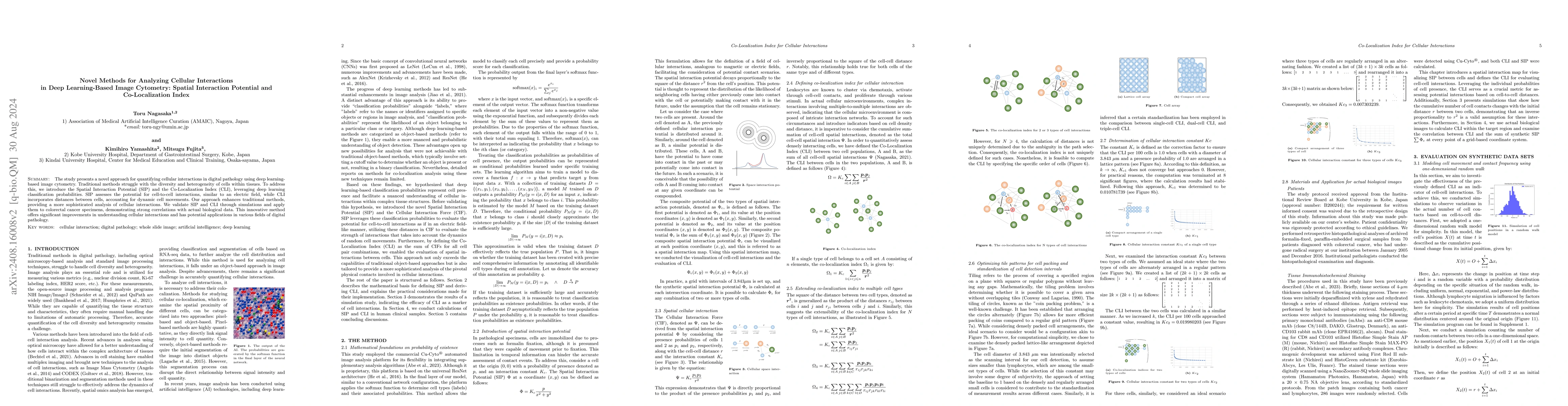
The study presents a novel approach for quantifying cellular interactions in digital pathology using deep learning-based image cytometry. Traditional methods struggle with the diversity and heterogene...

Thumbnail for Reliability Assessment Framework Based on Feature Separability for Pathological Cell Image Classification under Prior Bias

Reliability Assessment Framework Based on Feature Separability for Pathological Cell Image Classification under Prior Bias

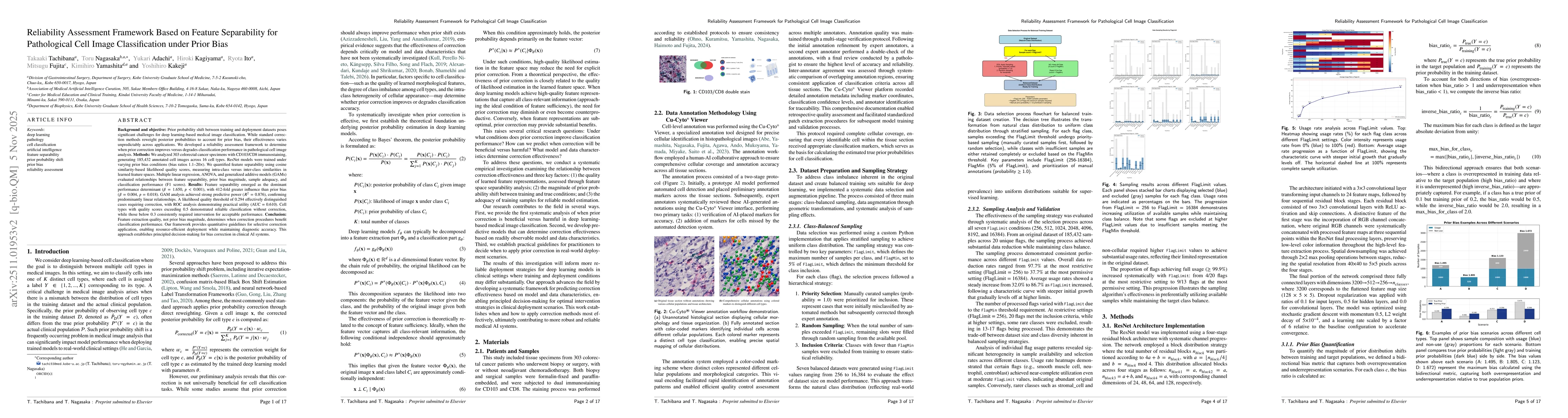
Background and objective: Prior probability shift between training and deployment datasets challenges deep learning-based medical image classification. Standard correction methods reweight posterior p...

Thumbnail for Spatially-extended Flow Phixer (SpeF-Phixer): A Spatially Extended $\varphi$-Mixing Framework for Gene Regulatory Causal Inference in Spatial Gene Field

Spatially-extended Flow Phixer (SpeF-Phixer): A Spatially Extended $\varphi$-Mixing Framework for Gene Regulatory Causal Inference in Spatial Gene Field

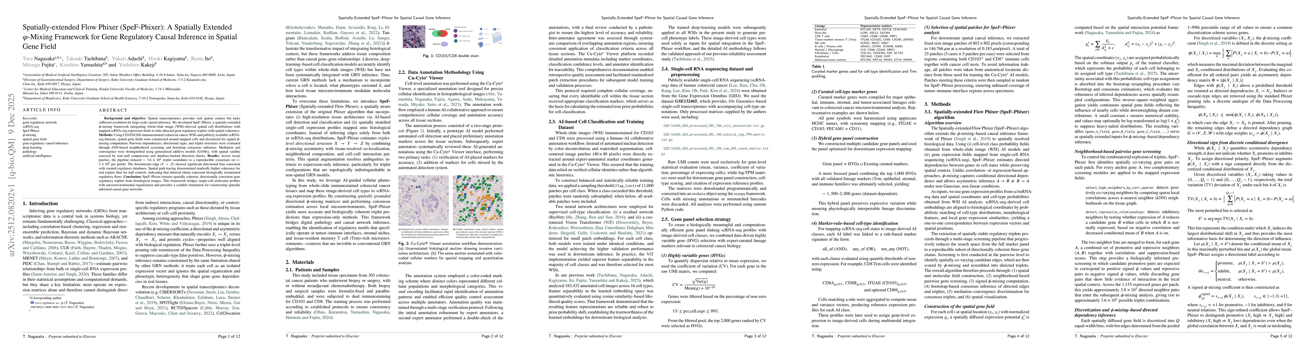
Background and objective: Spatial transcriptomics provides rich spatial context but lacks sufficient resolution for large-scale causal inference. We developed SpeF-Phixer, a spatially extended phi-mix...

Thumbnail for Revisiting the Role of Foundation Models in Cell-Level Histopathological Image Analysis under Small-Patch Constraints -- Effects of Training Data Scale and Blur Perturbations on CNNs and Vision Transformers

Revisiting the Role of Foundation Models in Cell-Level Histopathological Image Analysis under Small-Patch Constraints -- Effects of Training Data Scale and Blur Perturbations on CNNs and Vision Transformers

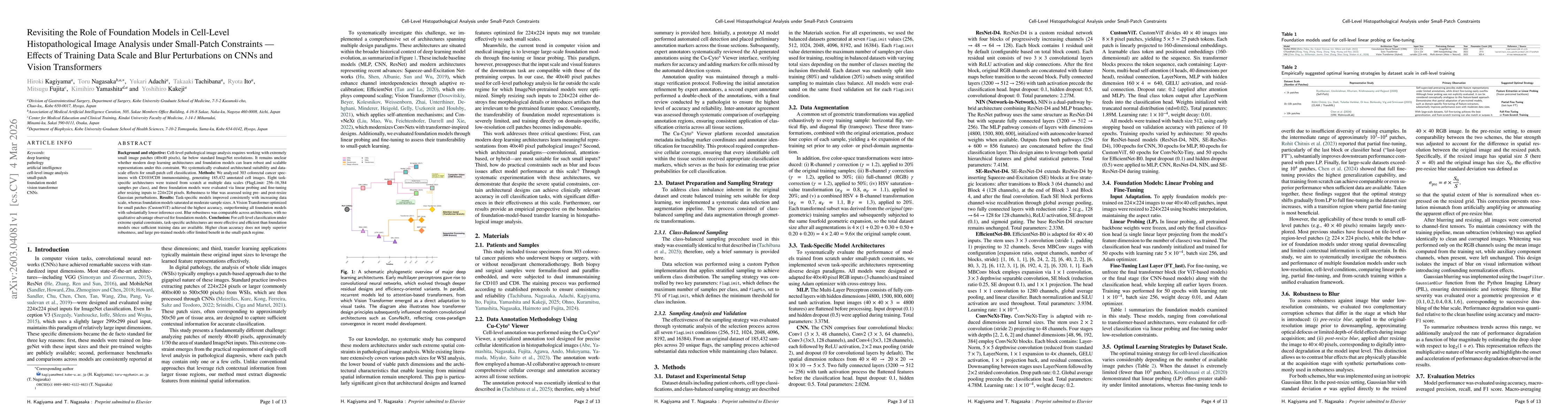
Background and objective: Cell-level pathological image analysis requires working with extremely small image patches (40x40 pixels), far below standard ImageNet resolutions. It remains unclear whether...

This website uses cookies to ensure you get the best experience. Learn more