Academic Profile
Statistics
Similar Authors
Papers on arXiv
Proton magnetic resonance spectroscopic imaging (1H-MRSI) is a powerful tool that enables the multidimensional non-invasive mapping of the neurochemical profile at high-resolution over the entire br...
Magnetic resonance spectroscopic imaging (MRSI) enables the simultaneous non-invasive acquisition of MR spectra from multiple spatial locations inside the brain. While 1H-MRSI is increasingly used i...
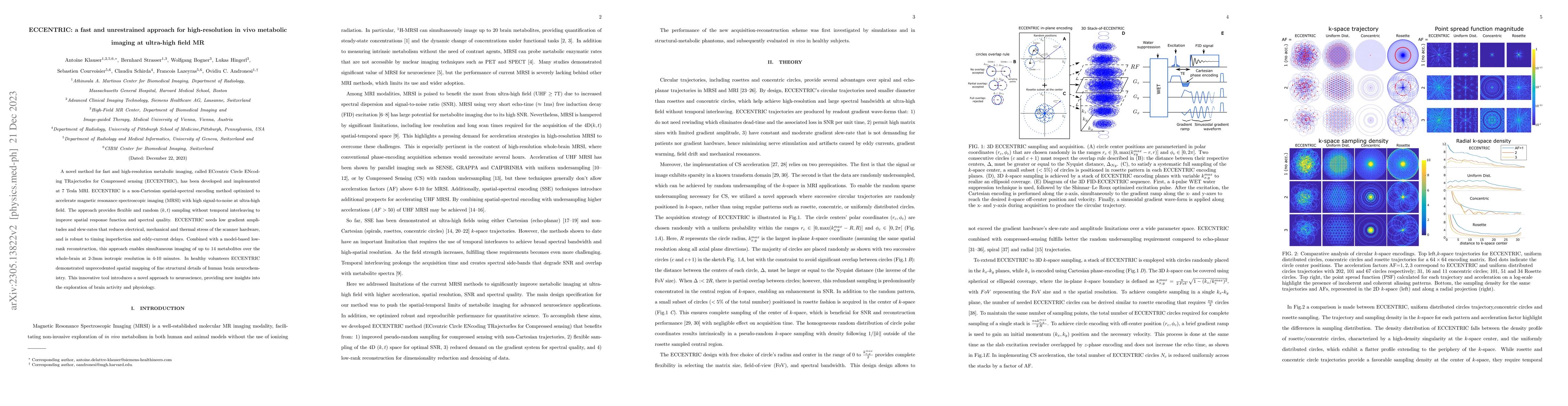
A novel method for fast and high-resolution metabolic imaging, called ECcentric Circle ENcoding TRajectorIes for Compressed sensing (ECCENTRIC), has been developed and implemented at 7 Tesla MRI. EC...
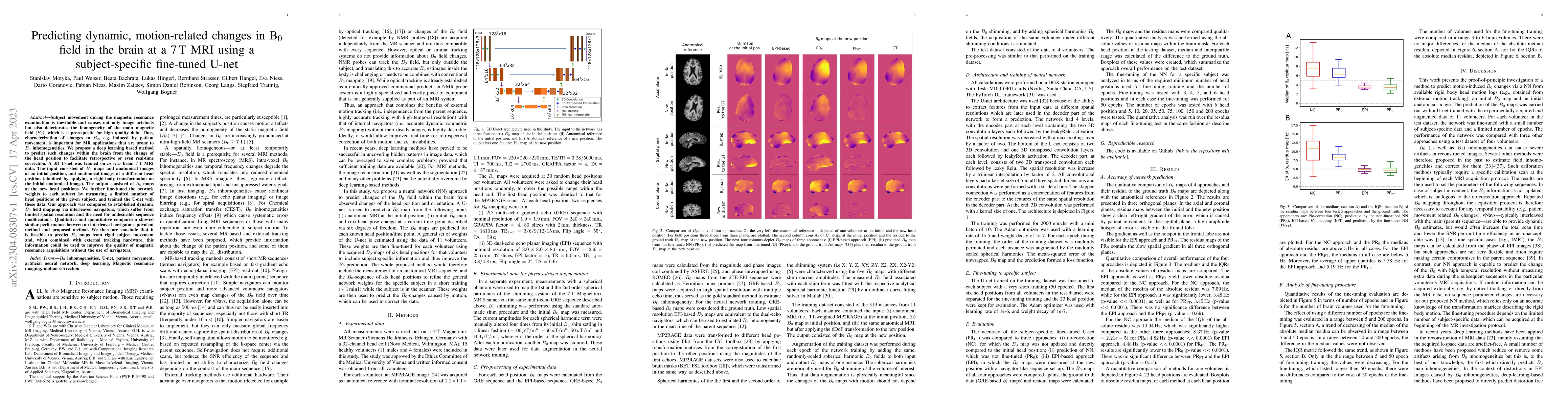
Subject movement during the magnetic resonance examination is inevitable and causes not only image artefacts but also deteriorates the homogeneity of the main magnetic field (B0), which is a prerequ...
This paper investigates the correlation between magnetic resonance spectroscopic imaging (MRSI) and magnetic resonance fingerprinting (MRF) in glioma patients by comparing neuro-oncological markers ...
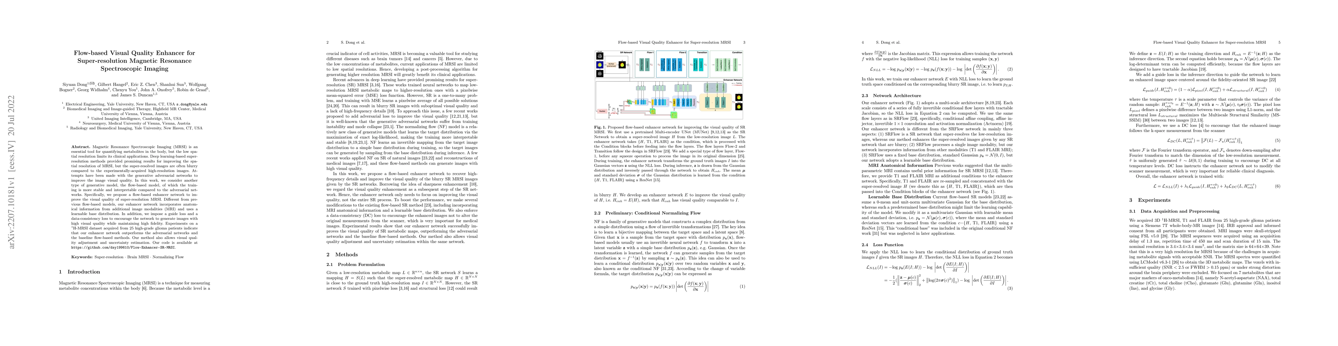
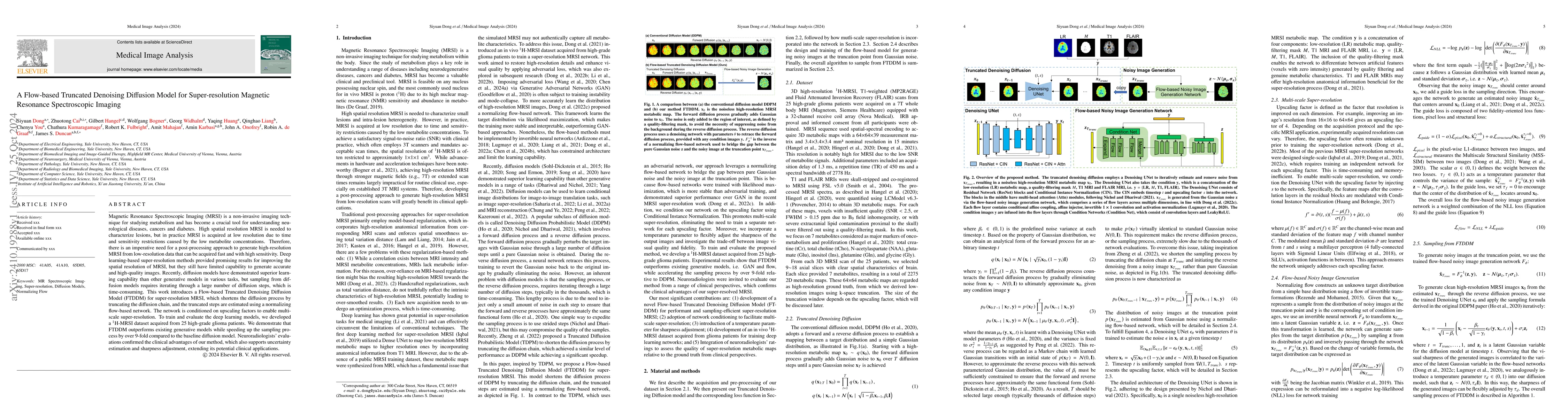
Magnetic Resonance Spectroscopic Imaging (MRSI) is an essential tool for quantifying metabolites in the body, but the low spatial resolution limits its clinical applications. Deep learning-based sup...
Magnetic Resonance Spectroscopic Imaging (MRSI) is a valuable tool for studying metabolic activities in the human body, but the current applications are limited to low spatial resolutions. The exist...
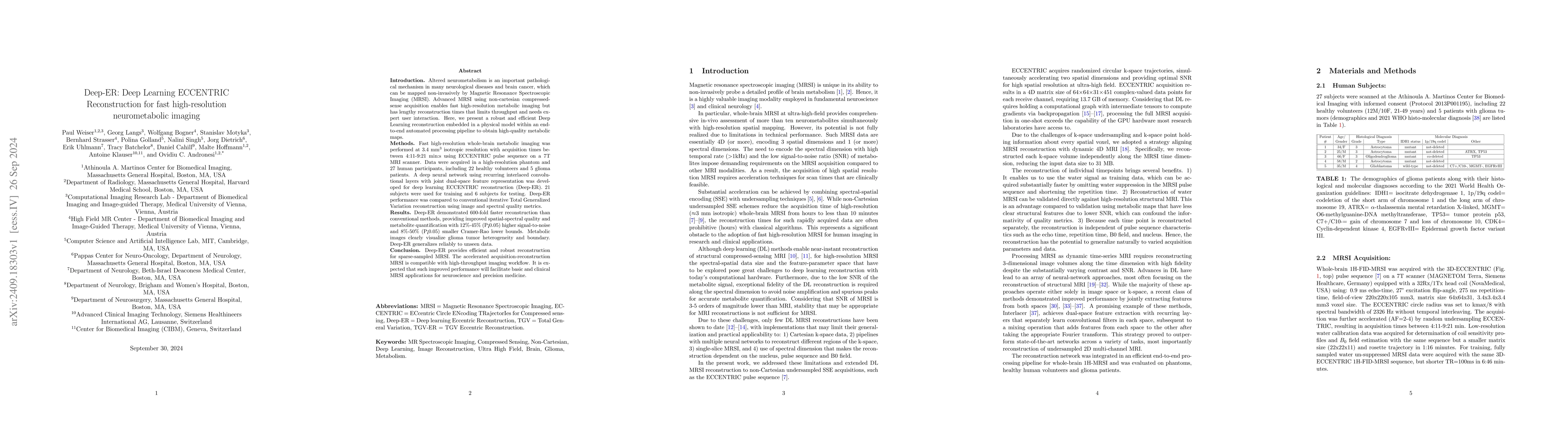
Introduction: Altered neurometabolism is an important pathological mechanism in many neurological diseases and brain cancer, which can be mapped non-invasively by Magnetic Resonance Spectroscopic Imag...
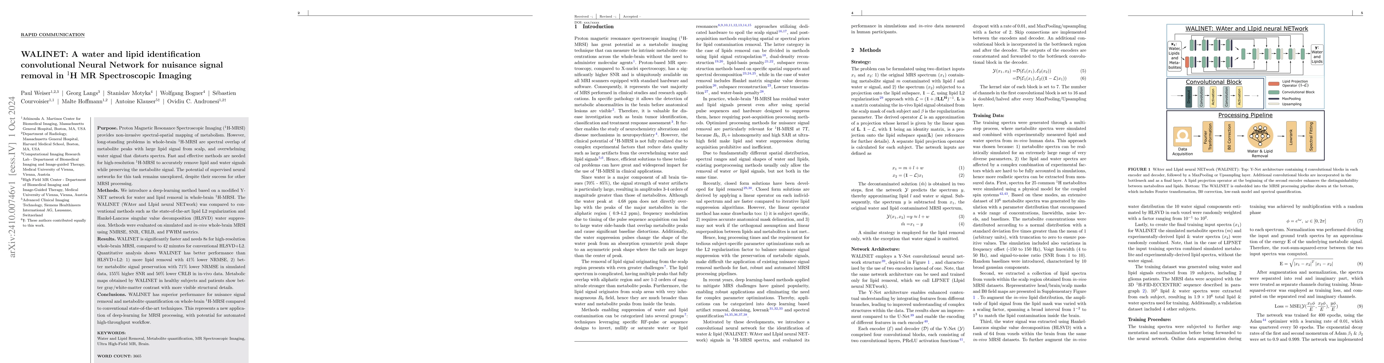
Purpose. Proton Magnetic Resonance Spectroscopic Imaging (1H-MRSI) provides non-invasive spectral-spatial mapping of metabolism. However, long-standing problems in whole-brain 1H-MRSI are spectral ove...
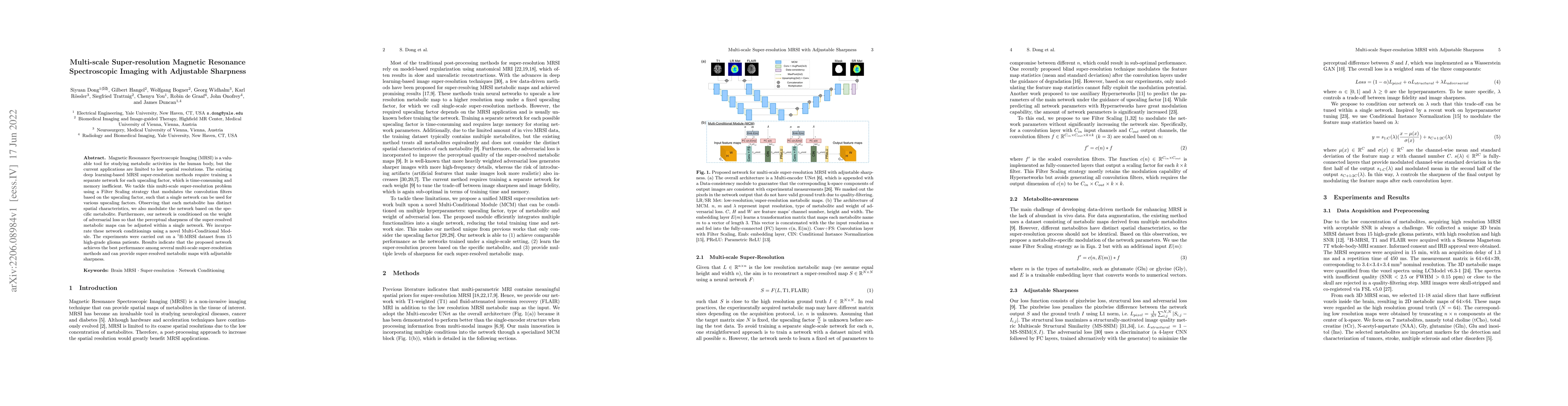
Magnetic Resonance Spectroscopic Imaging (MRSI) is a non-invasive imaging technique for studying metabolism and has become a crucial tool for understanding neurological diseases, cancers and diabetes....
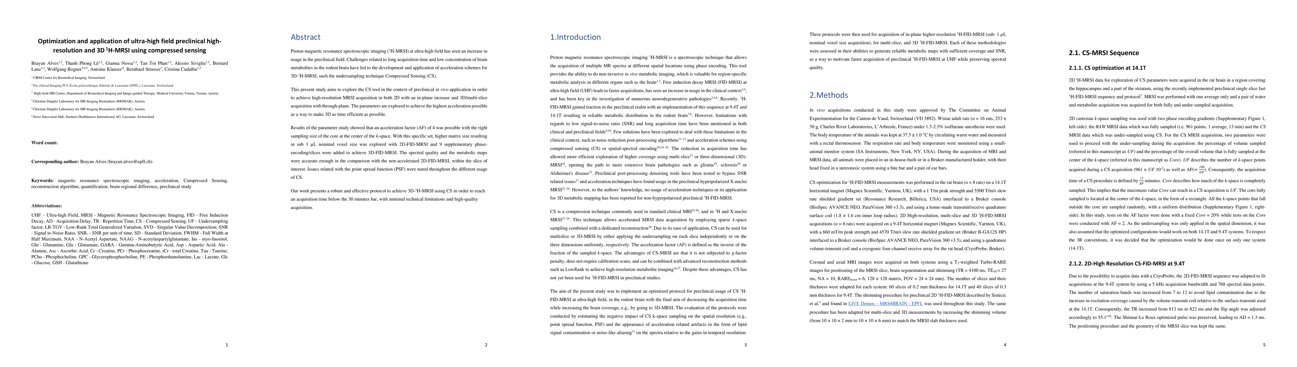
Proton magnetic resonance spectroscopic imaging (1H-MRSI) at ultra-high field has seen an increase in usage in the preclinical field. Challenges related to long acquisition time and low concentration ...
The use of synthetic data has emerged as an essential tool in Magnetic Resonance Spectroscopy (MRS) research and applications, providing advantages for optimization of acquisition, software validation...
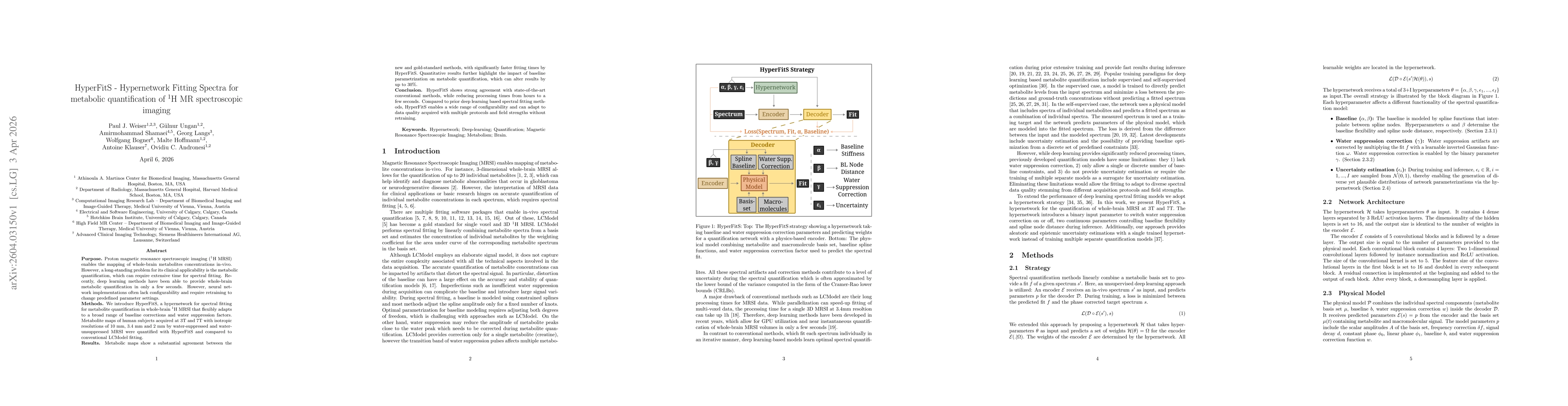
Purpose: Proton magnetic resonance spectroscopic imaging ($^1$H MRSI) enables the mapping of whole-brain metabolites concentrations in-vivo. However, a long-standing problem for its clinical applicabi...