Academic Profile
Statistics
Similar Authors
Papers on arXiv
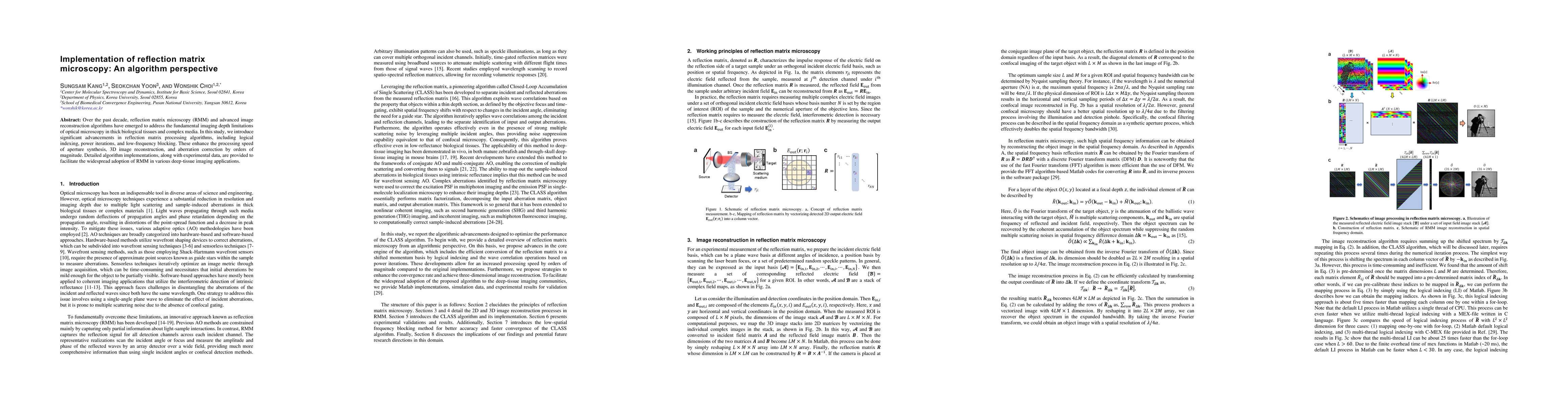
Over the past decade, reflection matrix microscopy (RMM) and advanced image reconstruction algorithms have emerged to address the fundamental imaging depth limitations of optical microscopy in thick...
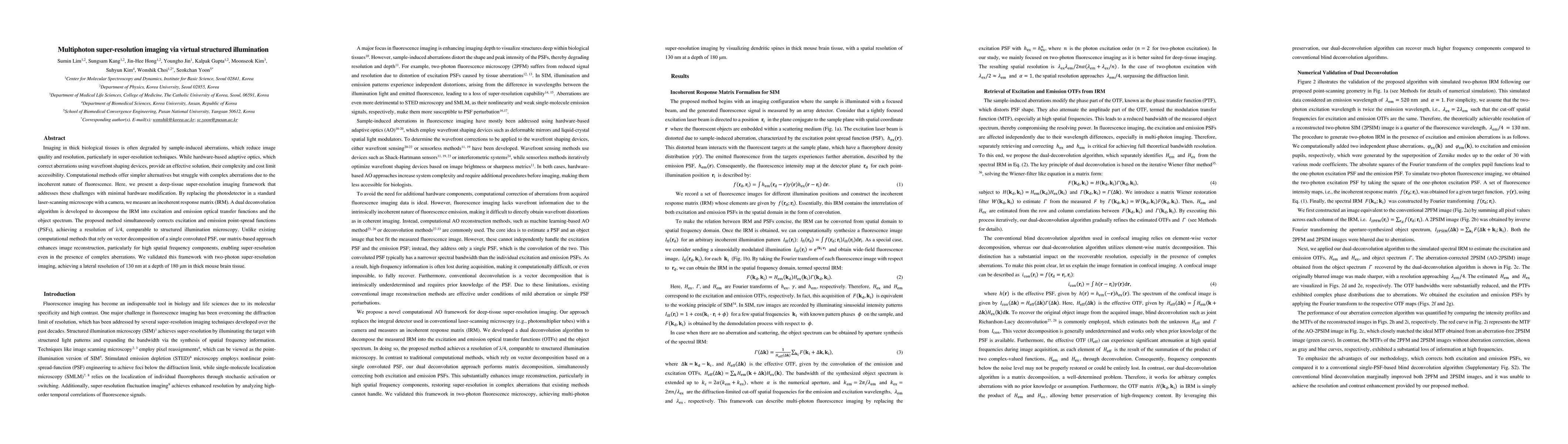
Fluorescence imaging in thick biological tissues is challenging due to sample-induced aberration and scattering, which leads to severe degradation of image quality and resolution. Fluorescence imagi...
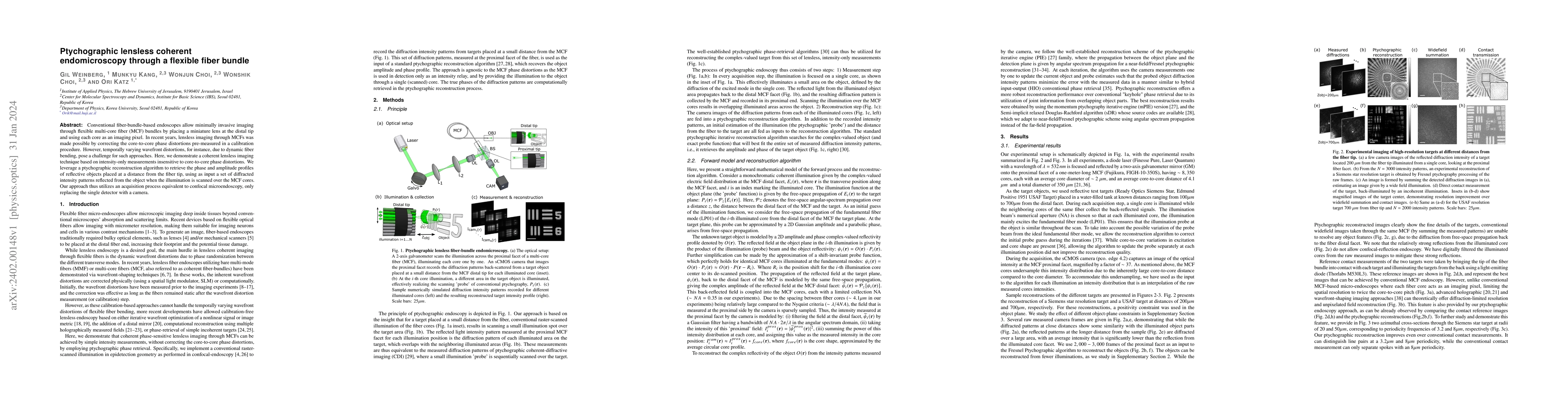
Conventional fiber-bundle-based endoscopes allow minimally invasive imaging through flexible multi-core fiber (MCF) bundles by placing a miniature lens at the distal tip and using each core as an im...
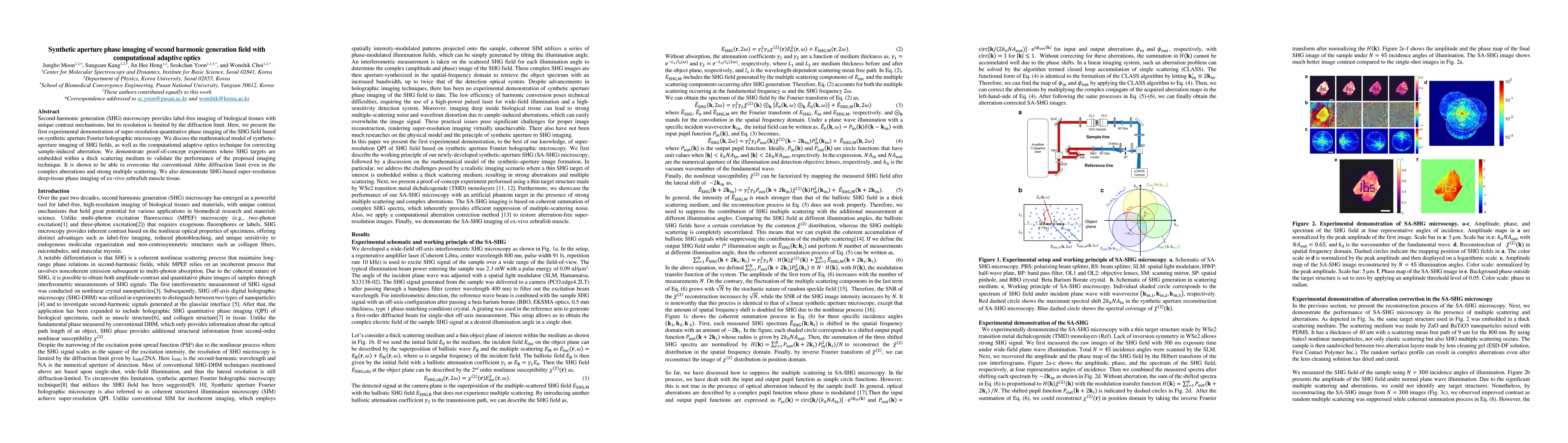
Second-harmonic generation (SHG) microscopy provides label-free imaging of biological tissues with unique contrast mechanisms, but its resolution is limited by the diffraction limit. Here, we presen...
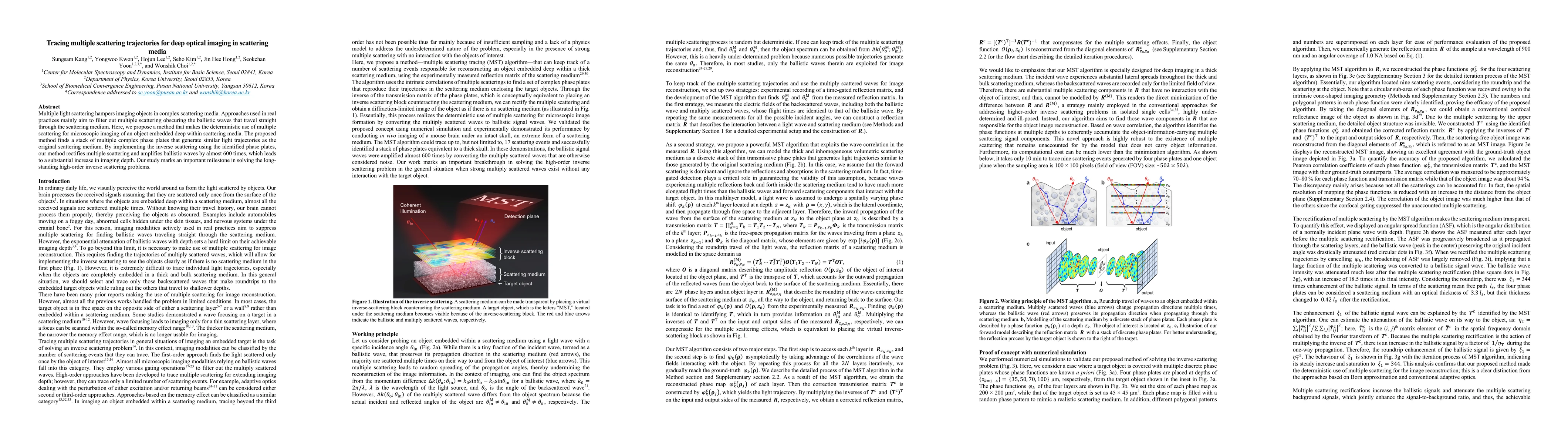
Multiple light scattering hampers imaging objects in complex scattering media. Approaches used in real practices mainly aim to filter out multiple scattering obscuring the ballistic waves that trave...
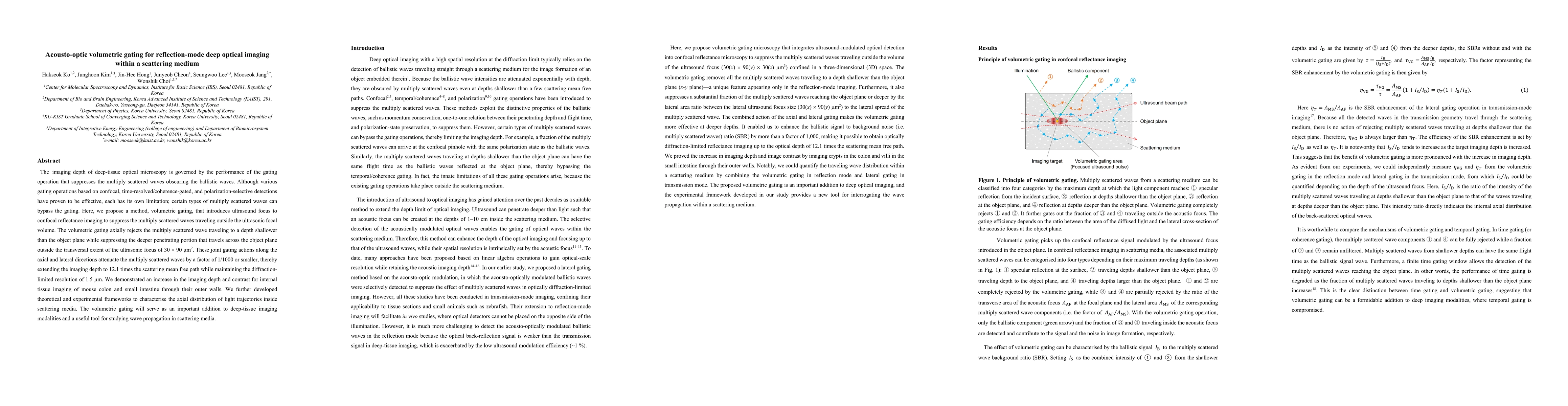
The imaging depth of deep-tissue optical microscopy is governed by the performance of the gating operation that suppresses the multiply scattered waves obscuring the ballistic waves. Although variou...
Imaging an object embedded within a scattering medium requires the correction of complex sample-induced wave distortions. Existing approaches have been designed to resolve them by optimizing signal ...
Shaping the wavefront of an incident wave to a complex scattering medium has demonstrated interesting possibilities, such as sub-diffraction wave focusing and enhancing light energy delivery. Howeve...
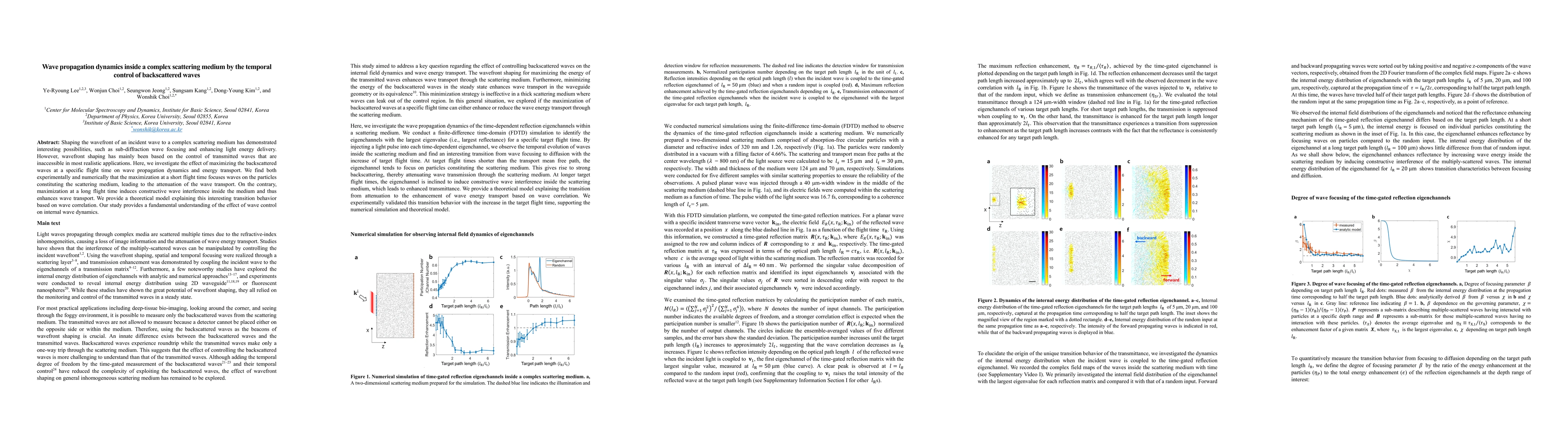
The last decade has seen the development of a wide set of tools, such as wavefront shaping, computational or fundamental methods, that allow to understand and control light propagation in a complex ...
Deep-tissue optical imaging suffers from the reduction of resolving power due to tissue-induced optical aberrations and multiple scattering noise. Reflection matrix approaches recording the maps of ...
Ultrathin lensless fibre endoscopes offer minimally invasive investigation, but they mostly operate as a rigid type due to the need for prior calibration of a fibre probe. Furthermore, most implemen...
We present a laser scanning reflection-matrix microscopy combining the scanning of laser focus and the wide-field mapping of the electric field of the backscattered waves for eliminating higher-orde...
Femtosecond-scale ultrafast imaging is an essential tool for visualizing ultrafast dynamics in molecular biology, physical chemistry, atomic physics, and fluid dynamics. Pump-probe imaging and a str...
Photoacoustic tomography (PAT) offers high optical contrast with acoustic imaging depth, making it essential for biomedical applications. While many all-optical systems have been developed to address ...
High-resolution optical microscopy has transformed biological imaging, yet its resolution and contrast deteriorate with depth due to multiple light scattering. Conventional correction strategies typic...
The refractive index (RI) is an intrinsic, label-free marker of a living cell's dry mass and subcellular morphology, and hence of its physiological state. Its three-dimensional (3D) reconstruction has...