Academic Profile
Statistics
Similar Authors
Papers on arXiv
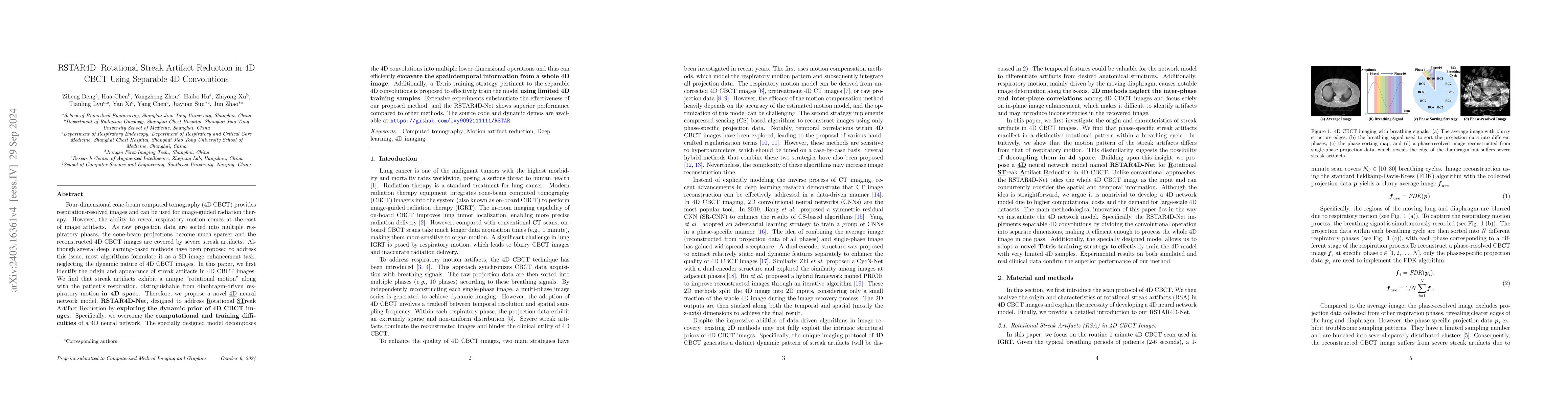
Four-dimensional cone-beam computed tomography (4D CBCT) provides respiration-resolved images and can be used for image-guided radiation therapy. However, the ability to reveal respiratory motion co...
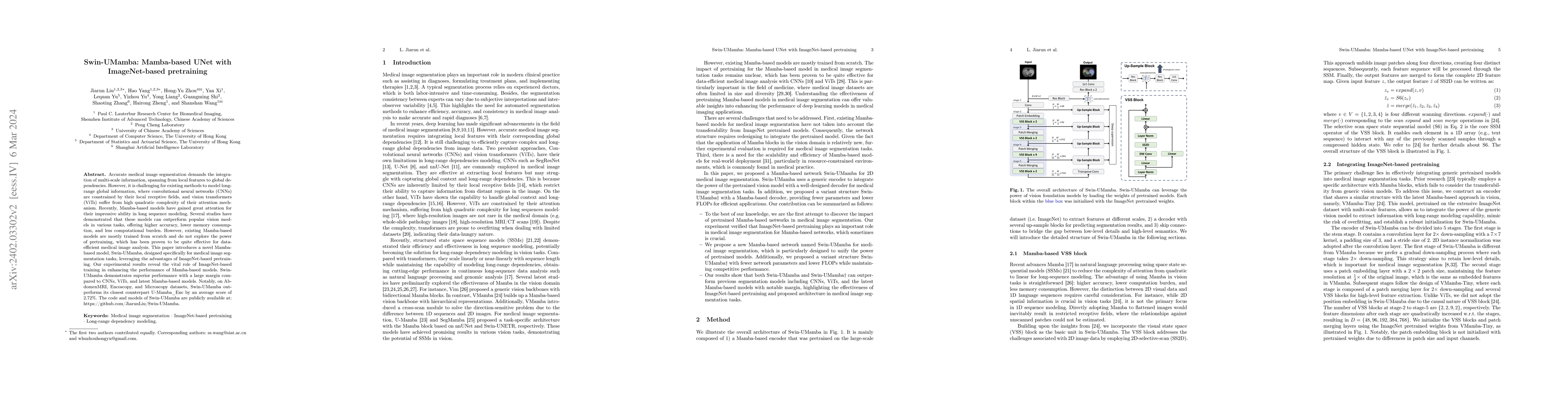
Accurate medical image segmentation demands the integration of multi-scale information, spanning from local features to global dependencies. However, it is challenging for existing methods to model ...
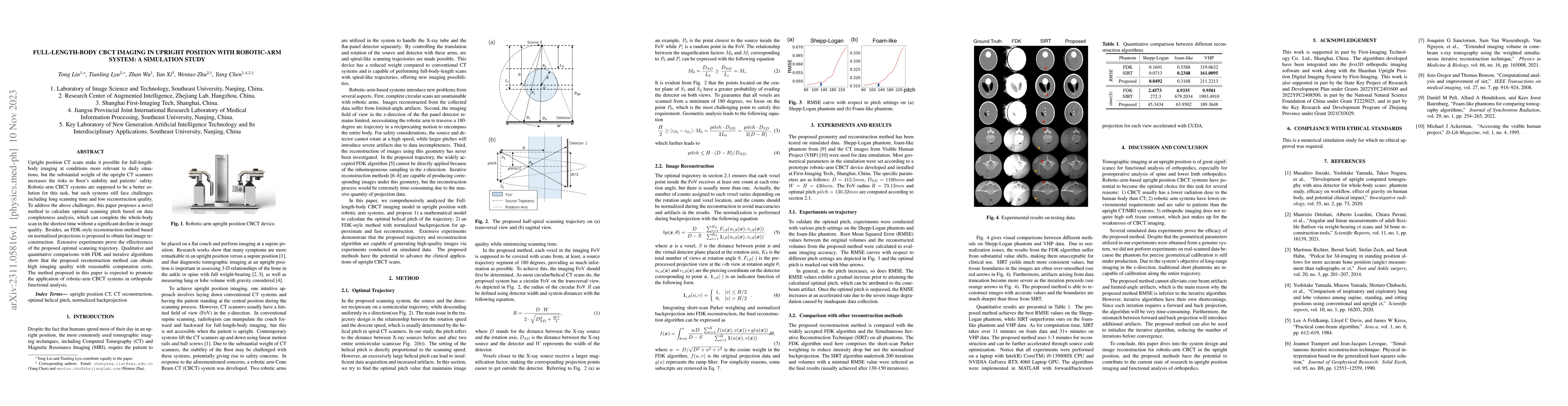
Upright position CT scans make it possible for full-length-body imaging at conditions more relevant to daily situations, but the substantial weight of the upright CT scanners increases the risks to ...
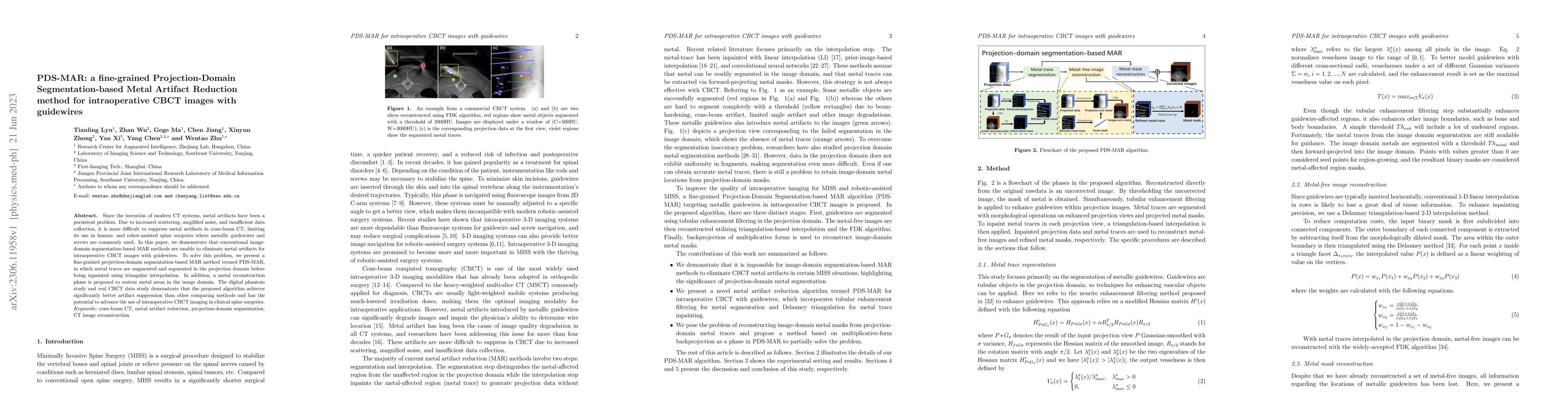
Since the invention of modern CT systems, metal artifacts have been a persistent problem. Due to increased scattering, amplified noise, and insufficient data collection, it is more difficult to supp...
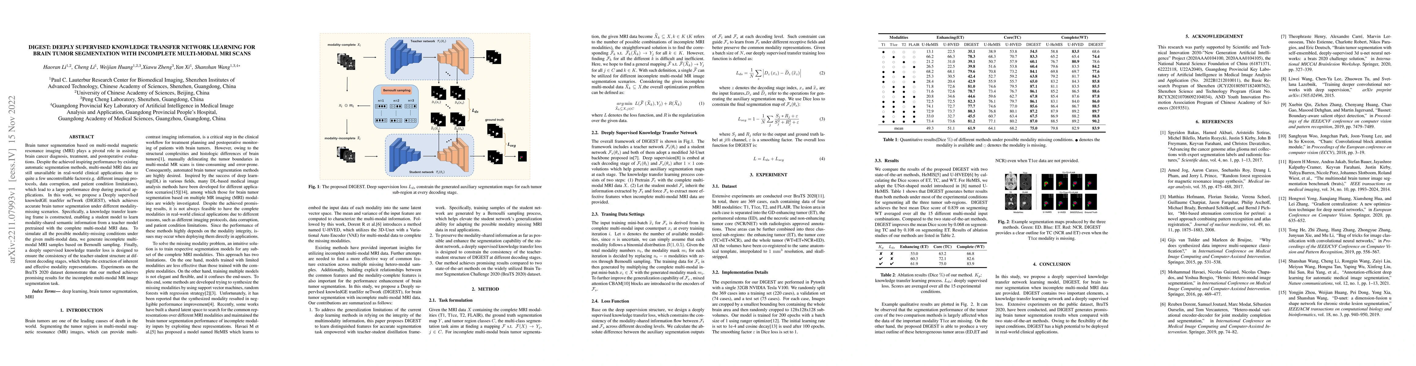
Brain tumor segmentation based on multi-modal magnetic resonance imaging (MRI) plays a pivotal role in assisting brain cancer diagnosis, treatment, and postoperative evaluations. Despite the achieve...
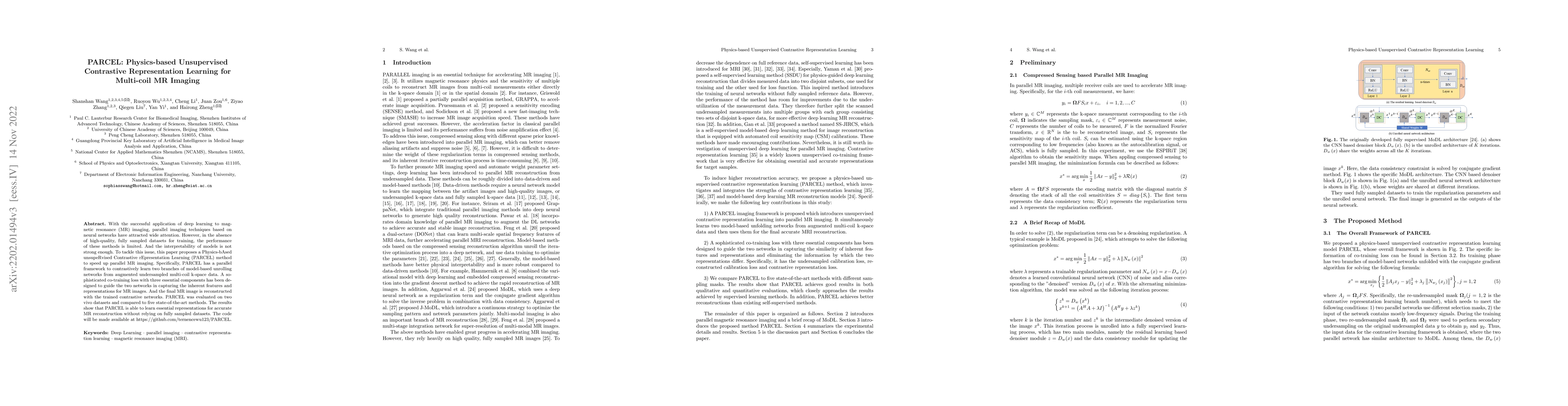
With the successful application of deep learning to magnetic resonance (MR) imaging, parallel imaging techniques based on neural networks have attracted wide attention. However, in the absence of hi...
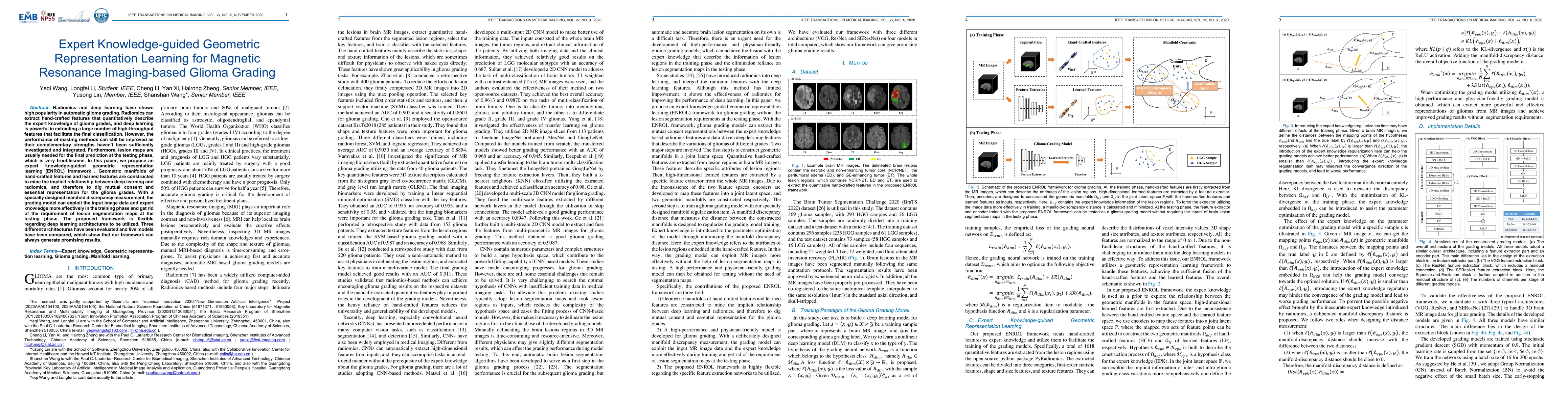
Radiomics and deep learning have shown high popularity in automatic glioma grading. Radiomics can extract hand-crafted features that quantitatively describe the expert knowledge of glioma grades, an...
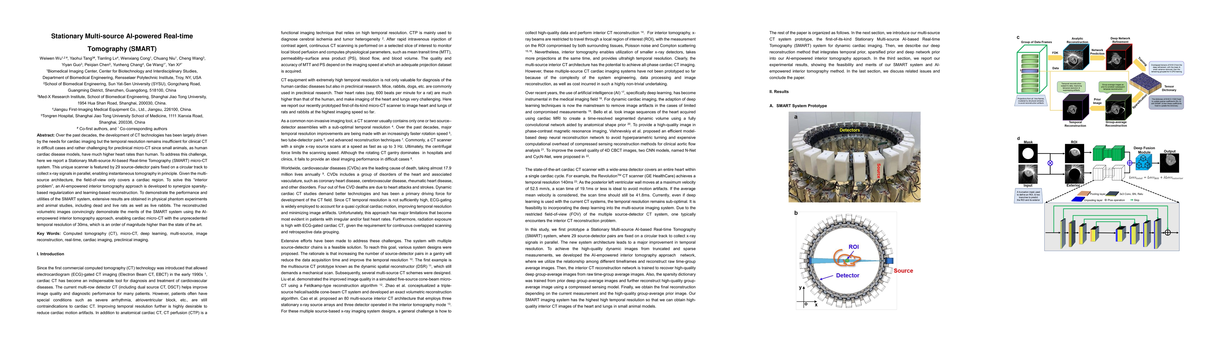
Over the past decades, the development of CT technologies has been largely driven by the needs for cardiac imaging but the temporal resolution remains insufficient for clinical CT in difficult cases...
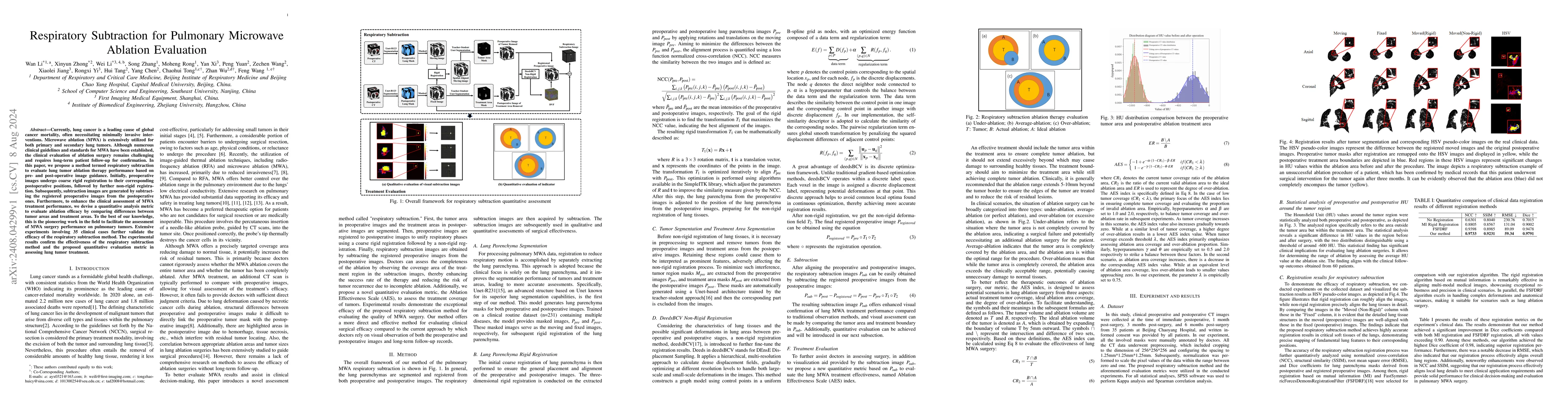
Currently, lung cancer is a leading cause of global cancer mortality, often necessitating minimally invasive interventions. Microwave ablation (MWA) is extensively utilized for both primary and second...
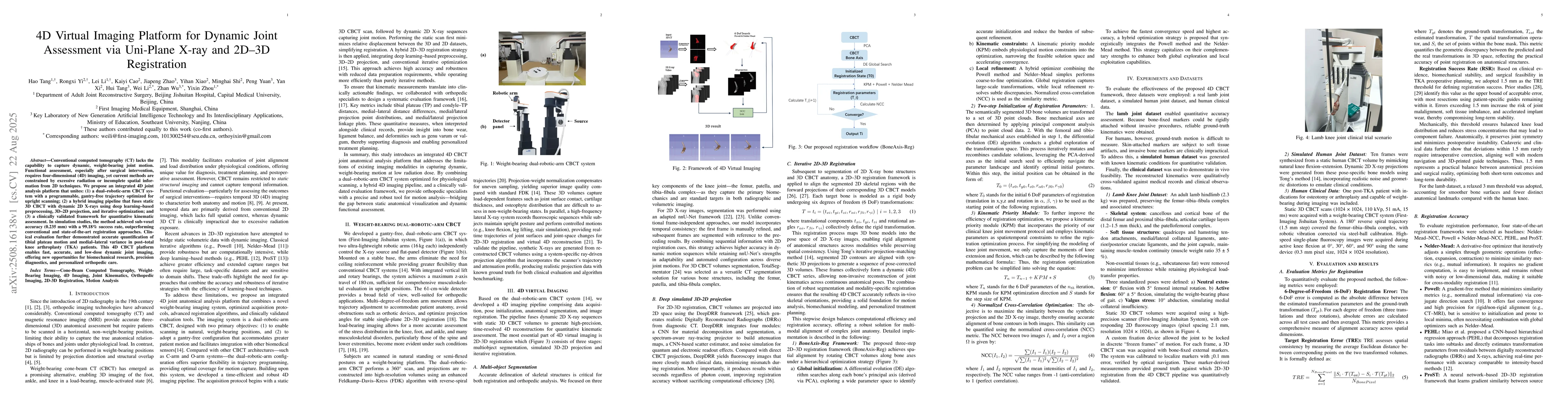
Conventional computed tomography (CT) lacks the ability to capture dynamic, weight-bearing joint motion. Functional evaluation, particularly after surgical intervention, requires four-dimensional (4D)...
Granular flows are ubiquitous in nature and industrial applications, yet a complete continuum theory remains a long-standing challenge. The leading empirical approach, μ(I) rheology, lacks microscopic...
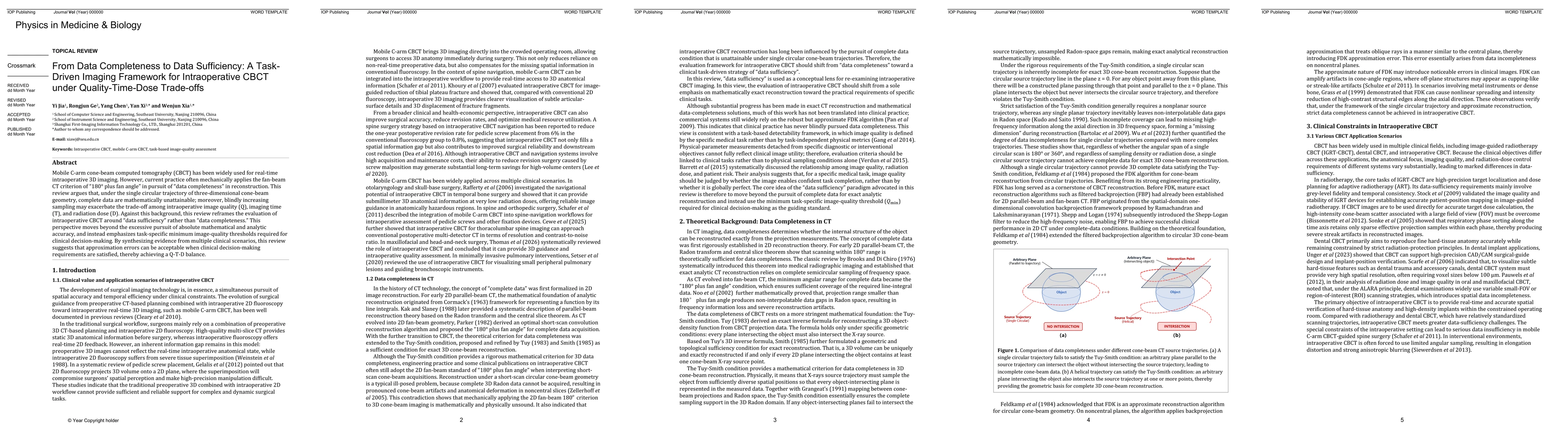
Mobile C-arm cone-beam computed tomography (CBCT) has been widely used for real-time intraoperative 3D imaging. However, current practice often mechanically applies the fan-beam CT criterion of "180° ...