Academic Profile
Statistics
Similar Authors
Papers on arXiv
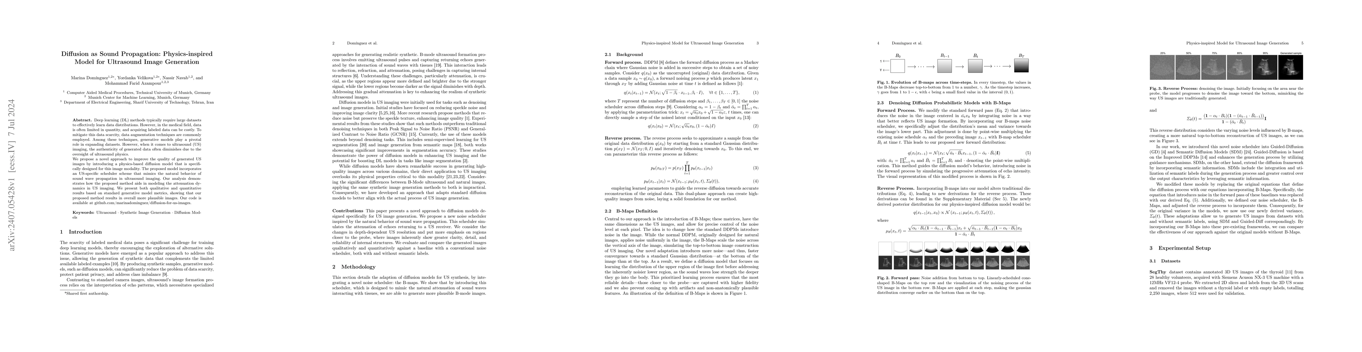
Deep learning (DL) methods typically require large datasets to effectively learn data distributions. However, in the medical field, data is often limited in quantity, and acquiring labeled data can be...
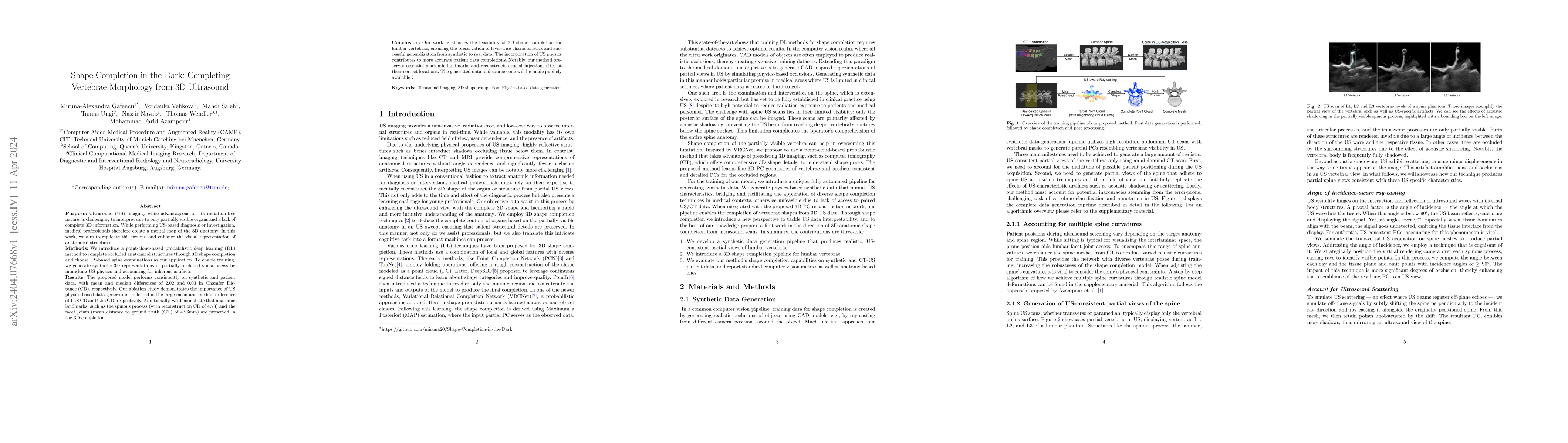
Purpose: Ultrasound (US) imaging, while advantageous for its radiation-free nature, is challenging to interpret due to only partially visible organs and a lack of complete 3D information. While perf...
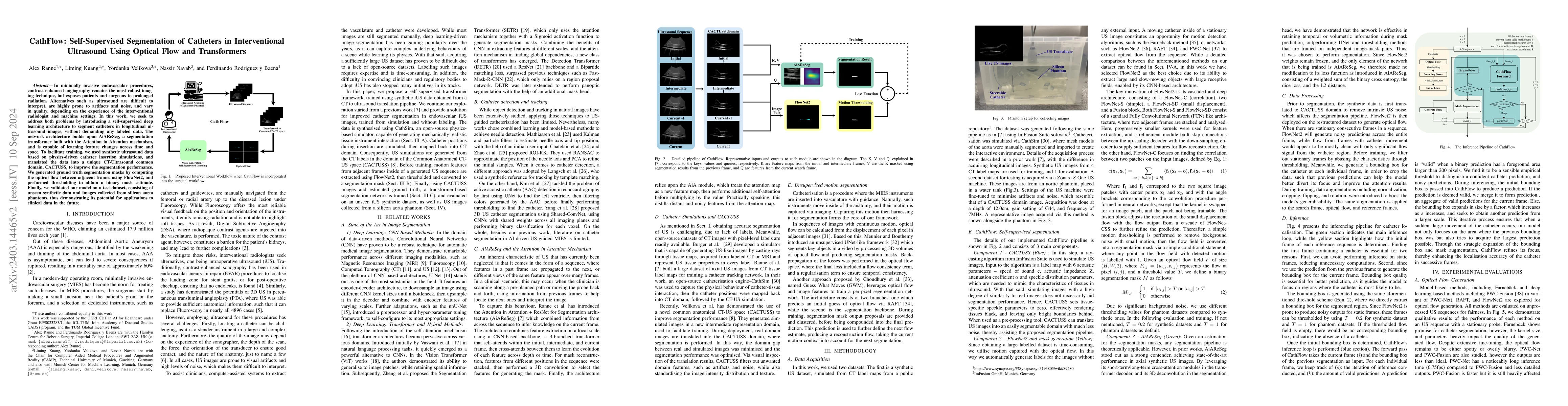
In minimally invasive endovascular procedures, contrast-enhanced angiography remains the most robust imaging technique. However, it is at the expense of the patient and clinician's health due to pro...
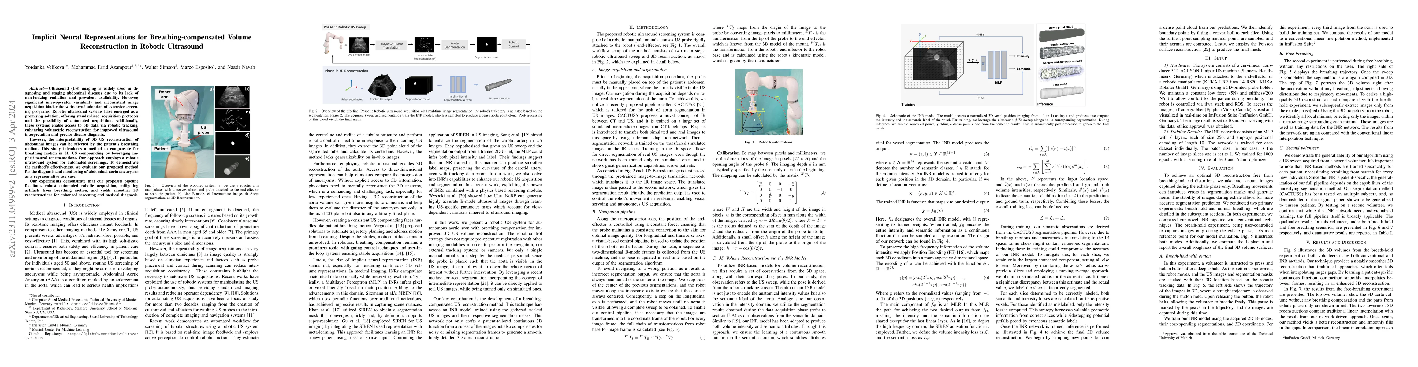
Ultrasound (US) imaging is widely used in diagnosing and staging abdominal diseases due to its lack of non-ionizing radiation and prevalent availability. However, significant inter-operator variabil...
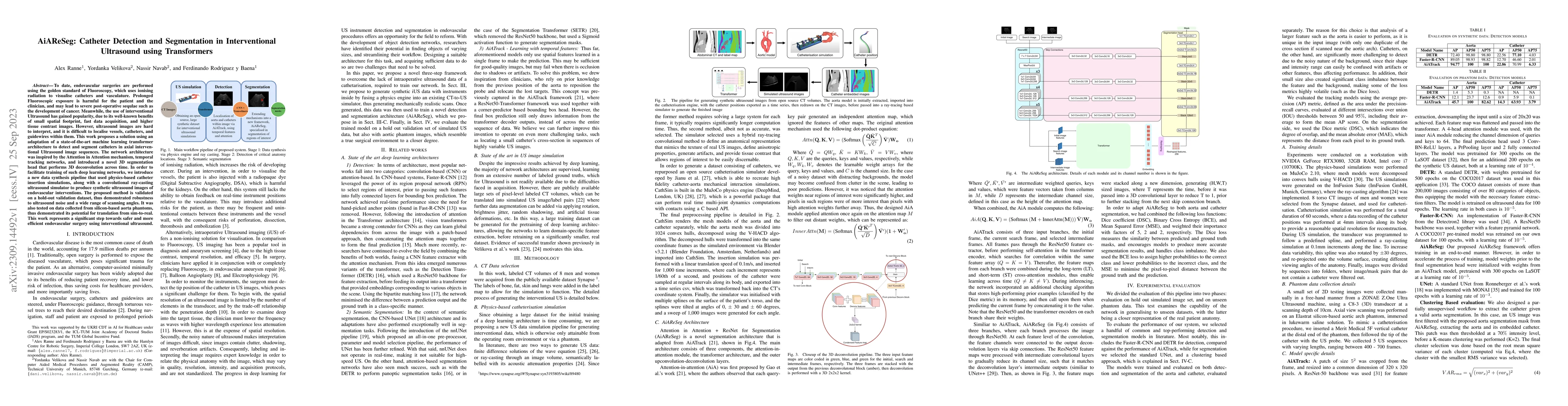
To date, endovascular surgeries are performed using the golden standard of Fluoroscopy, which uses ionising radiation to visualise catheters and vasculature. Prolonged Fluoroscopic exposure is harmf...
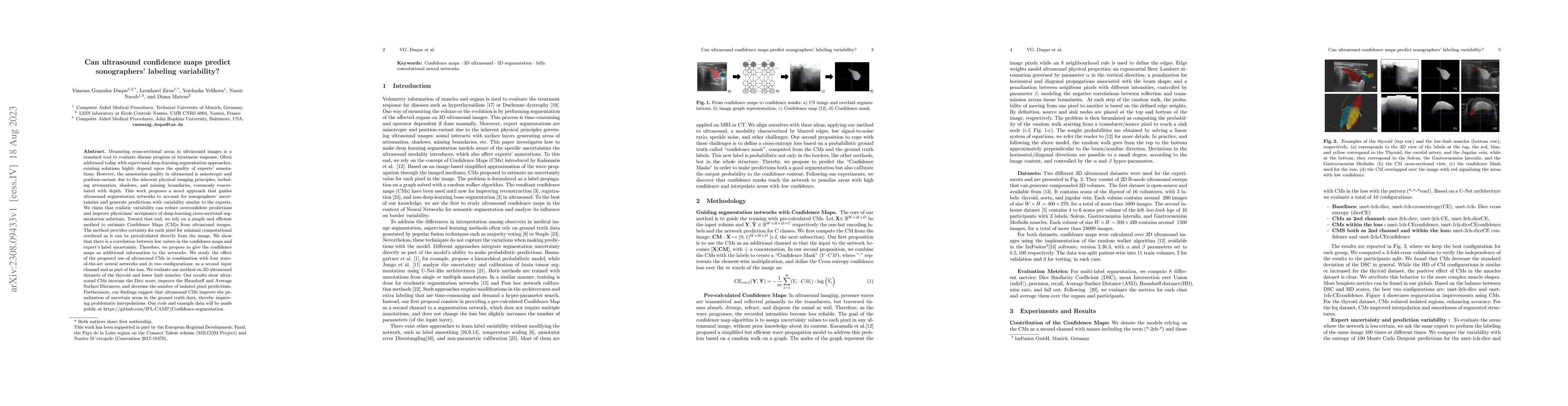
Measuring cross-sectional areas in ultrasound images is a standard tool to evaluate disease progress or treatment response. Often addressed today with supervised deep-learning segmentation approache...
Anatomical segmentation of organs in ultrasound images is essential to many clinical applications, particularly for diagnosis and monitoring. Existing deep neural networks require a large amount of ...
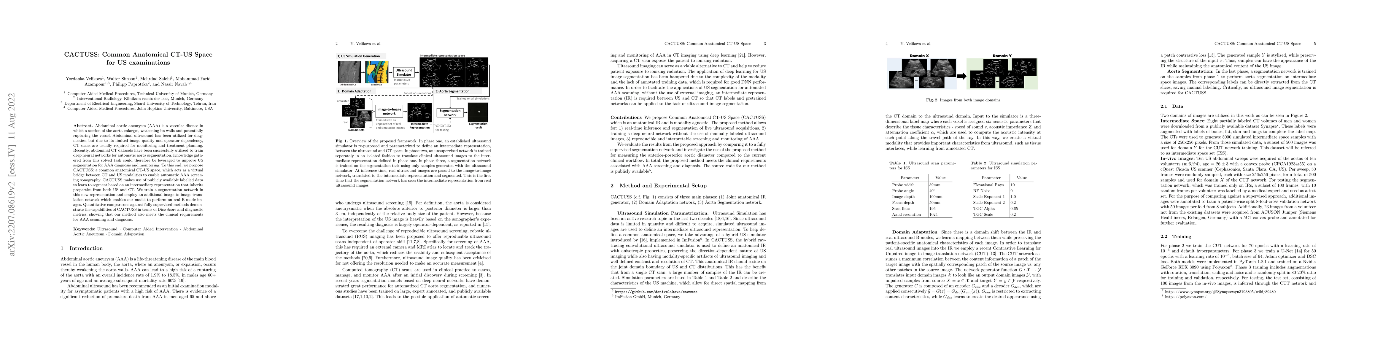
Abdominal aortic aneurysm (AAA) is a vascular disease in which a section of the aorta enlarges, weakening its walls and potentially rupturing the vessel. Abdominal ultrasound has been utilized for d...
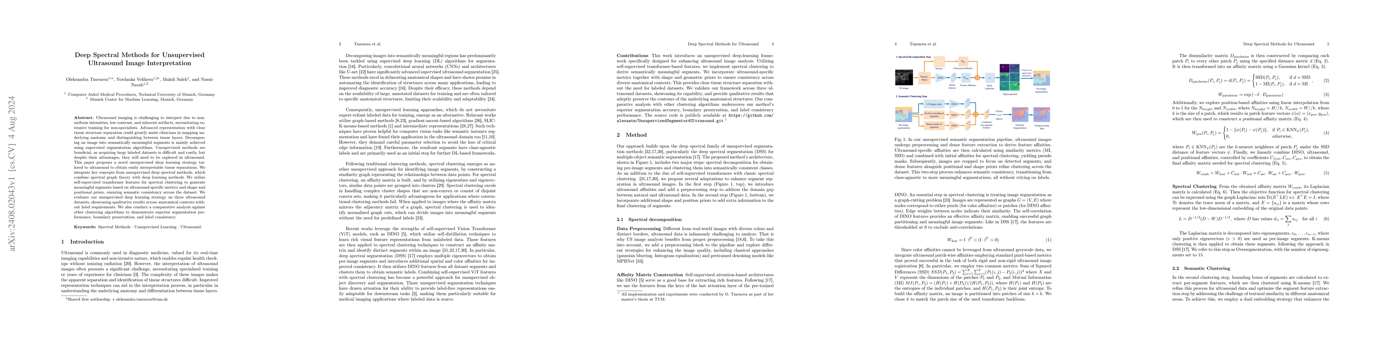
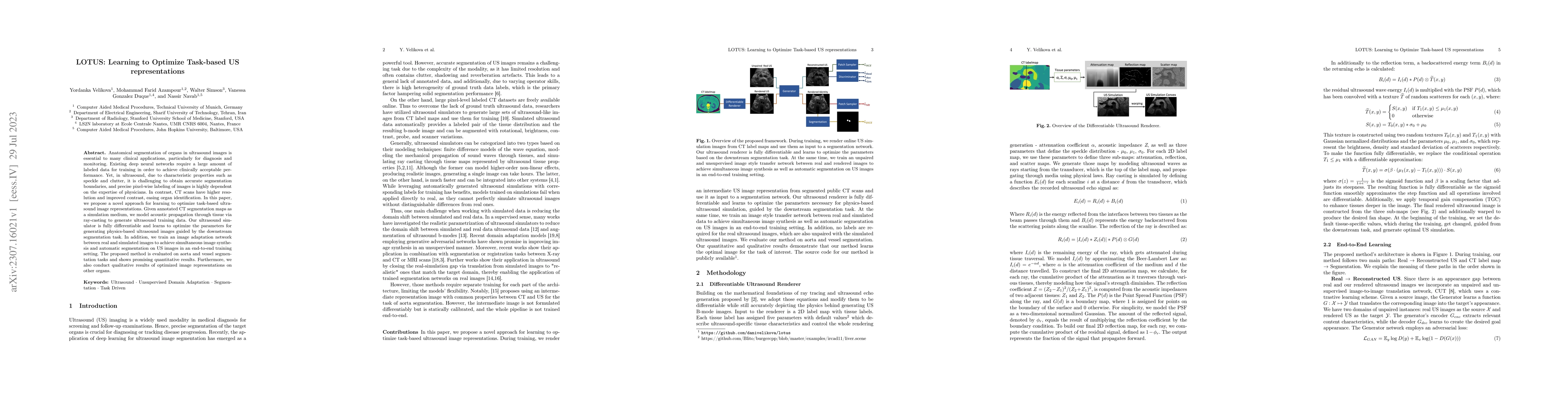
Ultrasound imaging is challenging to interpret due to non-uniform intensities, low contrast, and inherent artifacts, necessitating extensive training for non-specialists. Advanced representation with ...
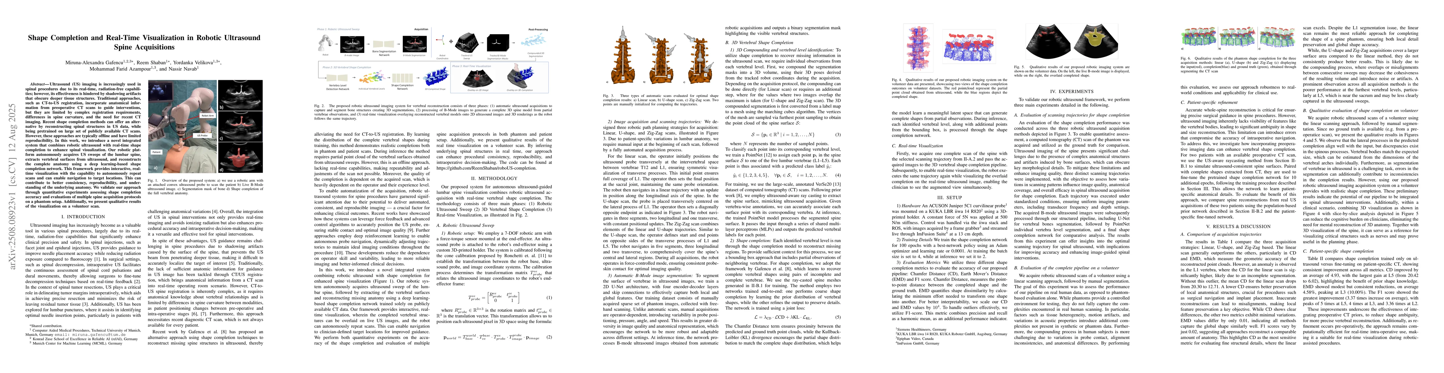
Ultrasound (US) imaging is increasingly used in spinal procedures due to its real-time, radiation-free capabilities; however, its effectiveness is hindered by shadowing artifacts that obscure deeper t...
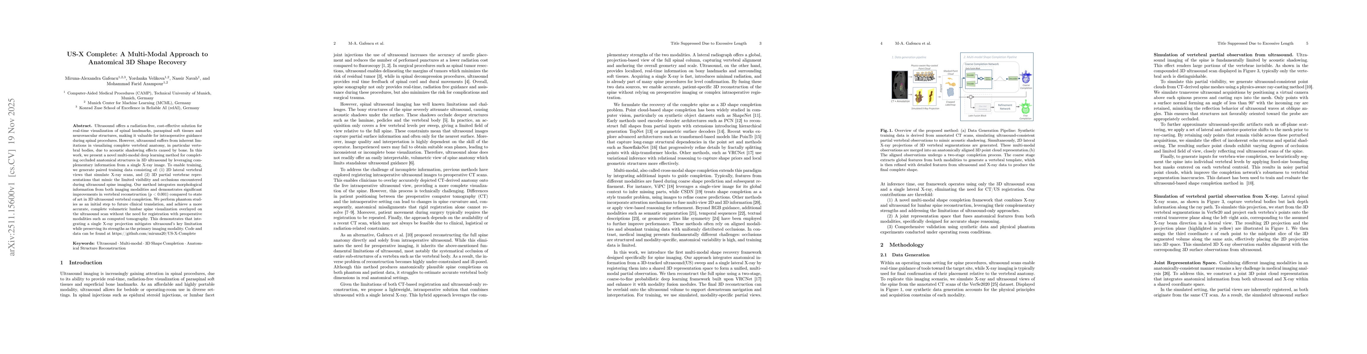
Ultrasound offers a radiation-free, cost-effective solution for real-time visualization of spinal landmarks, paraspinal soft tissues and neurovascular structures, making it valuable for intraoperative...
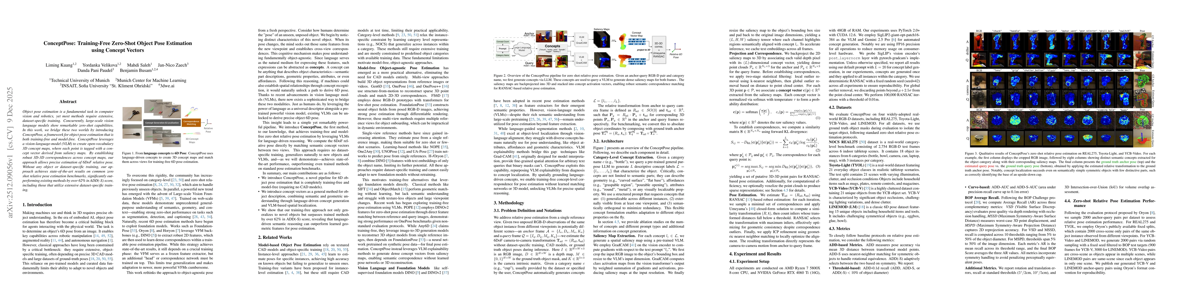
Object pose estimation is a fundamental task in computer vision and robotics, yet most methods require extensive, dataset-specific training. Concurrently, large-scale vision language models show remar...
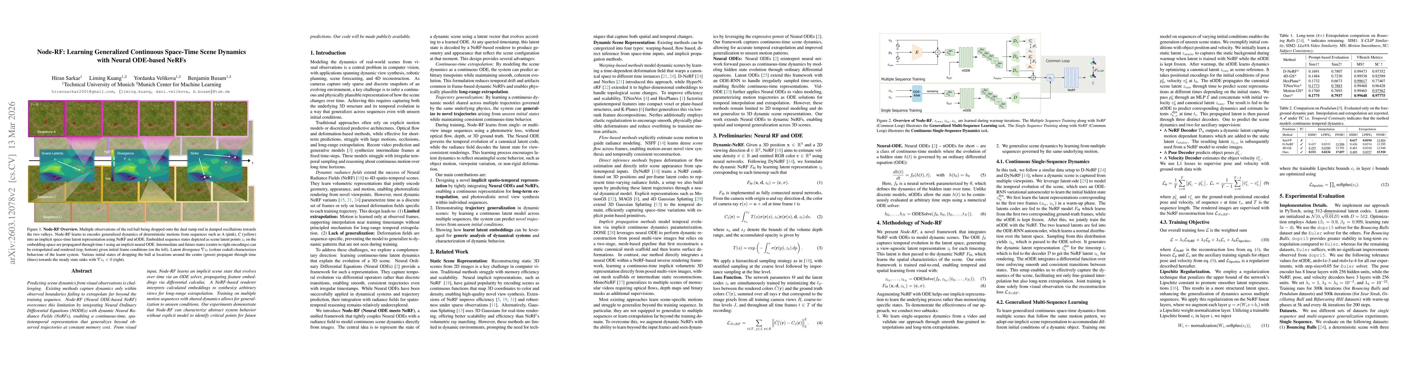
Predicting scene dynamics from visual observations is challenging. Existing methods capture dynamics only within observed boundaries failing to extrapolate far beyond the training sequence. Node-RF (N...
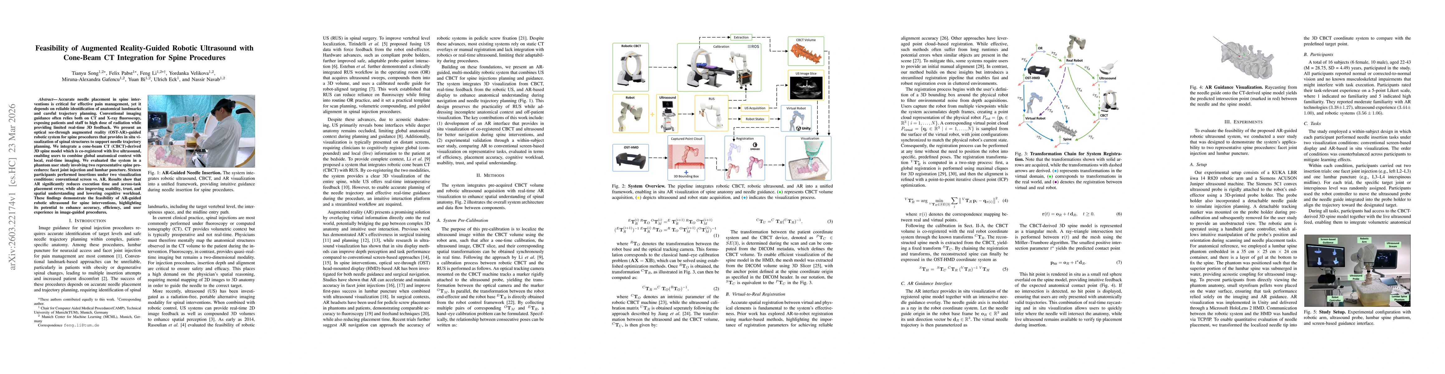
Accurate needle placement in spine interventions is critical for effective pain management, yet it depends on reliable identification of anatomical landmarks and careful trajectory planning. Conventio...
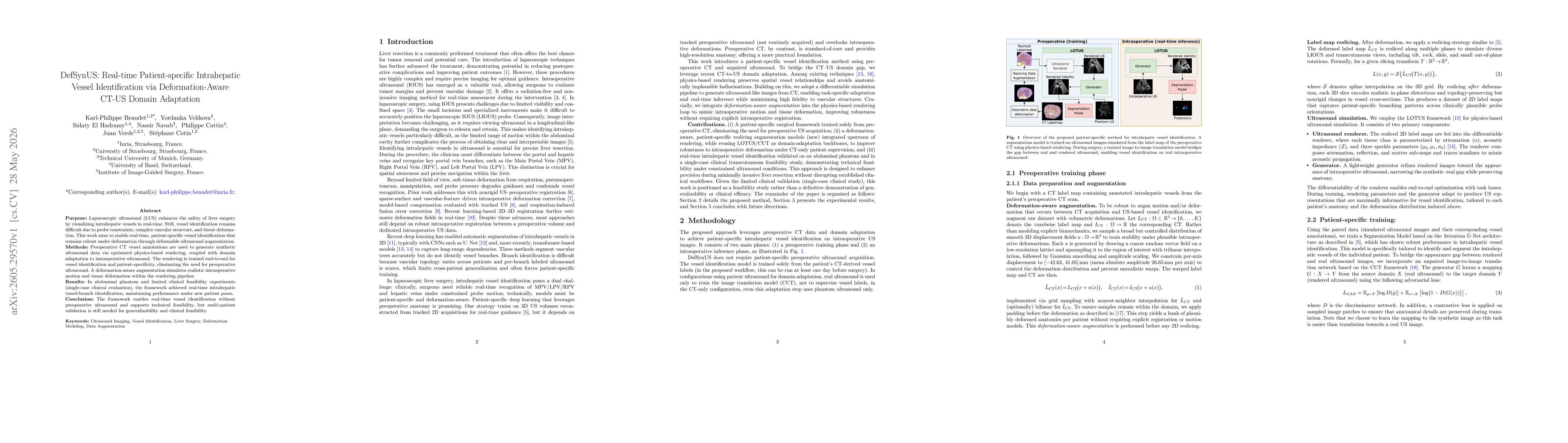
Purpose: Laparoscopic ultrasound (LUS) enhances the safety of liver surgery by visualizing intrahepatic vessels in real-time. Still, vessel identification remains difficult due to probe constraints, c...