2.5D Thermometry Maps for MRI-guided Tumor Ablation
Publication
Metrics
AI Quick Summary
Researchers developed a method to generate 2D thermometry maps from MRI phase images for accurate heat distribution monitoring during tumor ablation, with promising results including high accuracy and fast reconstruction times.
Paper Preview
Abstract
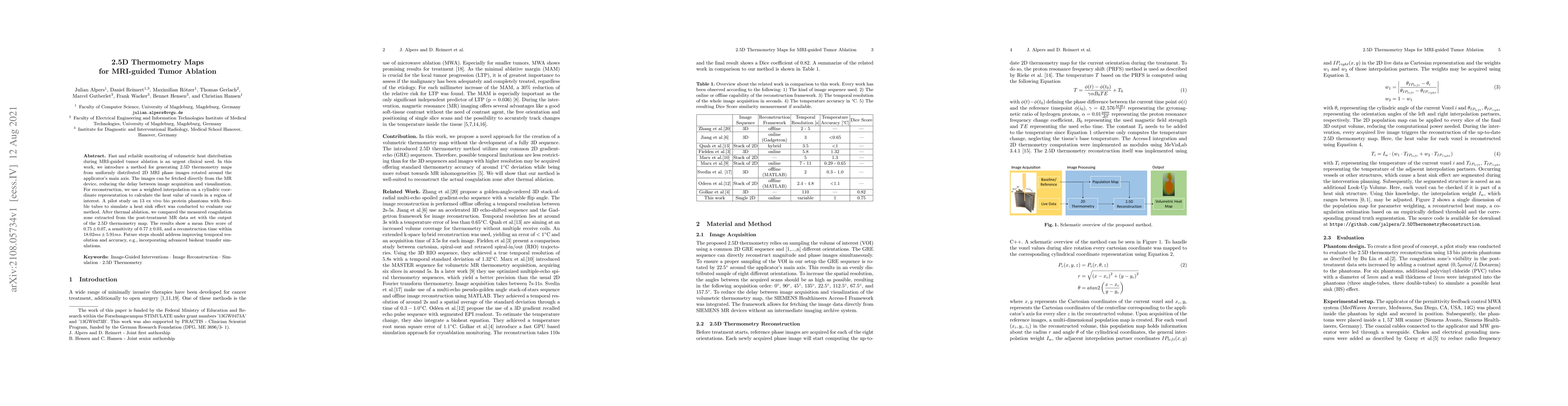
Fast and reliable monitoring of volumetric heat distribution during MRI-guided tumor ablation is an urgent clinical need. In this work, we introduce a method for generating 2.5D thermometry maps from uniformly distributed 2D MRI phase images rotated around the applicator's main axis. The images can be fetched directly from the MR device, reducing the delay between image acquisition and visualization. For reconstruction, we use a weighted interpolation on a cylindric coordinate representation to calculate the heat value of voxels in a region of interest. A pilot study on 13 ex vivo bio protein phantoms with flexible tubes to simulate a heat sink effect was conducted to evaluate our method. After thermal ablation, we compared the measured coagulation zone extracted from the post-treatment MR data set with the output of the 2.5D thermometry map. The results show a mean Dice score of 0.75+-0.07, a sensitivity of 0.77+-0.03, and a reconstruction time within 18.02ms+-5.91ms. Future steps should address improving temporal resolution and accuracy, e.g., incorporating advanced bioheat transfer simulations.
AI Key Findings
Get AI-generated insights about this paper's methodology, results, significance, and more — seven facets brought into focus.
Impact
Paper Details
Authors
PDF Preview
Key Terms
Citation Network
Current paper (gray), citations (green), references (blue)
Display is limited for performance on very large graphs.
Discussion 0