2D-Densely Connected Convolution Neural Networks for automatic Liver and Tumor Segmentation
Publication
Metrics
AI Quick Summary
This paper presents a two-stage DenseNet-based CNN for automatic liver and tumor segmentation in CT images. The liver and tumor models are independently trained and cascaded, with the liver model achieving a global dice score of 0.923 and the tumor model 0.625, along with an rmse of 0.044 for tumor burden.
Paper Preview
Abstract
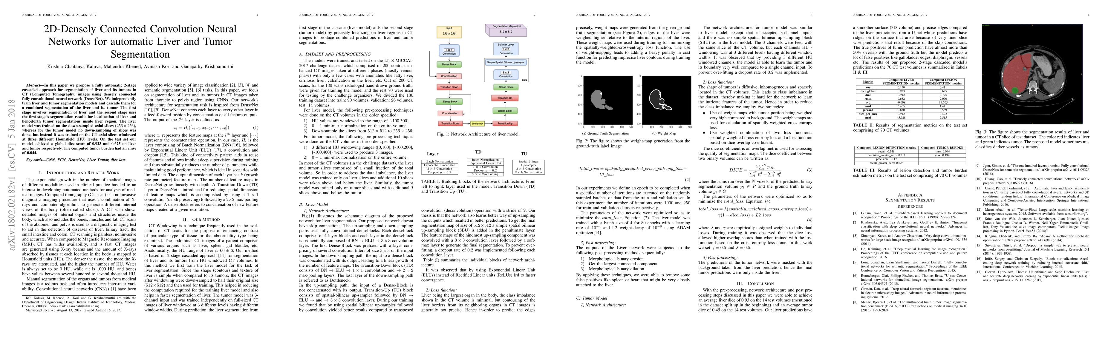
In this paper we propose a fully automatic 2-stage cascaded approach for segmentation of liver and its tumors in CT (Computed Tomography) images using densely connected fully convolutional neural network (DenseNet). We independently train liver and tumor segmentation models and cascade them for a combined segmentation of the liver and its tumor. The first stage involves segmentation of liver and the second stage uses the first stage's segmentation results for localization of liver and henceforth tumor segmentations inside liver region. The liver model was trained on the down-sampled axial slices $(256 \times 256)$, whereas for the tumor model no down-sampling of slices was done, but instead it was trained on the CT axial slices windowed at three different Hounsfield (HU) levels. On the test set our model achieved a global dice score of 0.923 and 0.625 on liver and tumor respectively. The computed tumor burden had an rmse of 0.044.
OpenAlex
- Citations in OpenAlex
- 68
- Publication year
- 2018
- Access status
- Open access
Source: OpenAlex · Snapshot
AI Key Findings
Get AI-generated insights about this paper's methodology, results, significance, and more — seven facets brought into focus.
Impact
Paper Details
PDF Preview
Key Terms
Citation Network
Current paper (gray), citations (green), references (blue)
Display is limited for performance on very large graphs.
Discussion 0