Quick Summary:

This paper proposes a two-step adversarial network for 3-D image-to-image fusion in lightsheet microscopy, addressing challenges of high resolution and global context. The method shows significant improvement in fusion quality, with average SSIM values of over 0.85 for nuclei and 0.91 for membranes, demonstrating potential to enhance lightsheet microscopy capabilities.

Summary

Lightsheet microscopy is a powerful 3-D imaging technique that addresses limitations of traditional optical and confocal microscopy but suffers from a low penetration depth and reduced image quality at greater depths. Multiview lightsheet microscopy improves 3-D resolution by combining multiple views but simultaneously increasing the complexity and the photon budget, leading to potential photobleaching and phototoxicity. The FuseMyCells challenge, organized in conjunction with the IEEE ISBI 2025 conference, aims to benchmark deep learning-based solutions for fusing high-quality 3-D volumes from single 3-D views, potentially simplifying procedures and conserving the photon budget. 
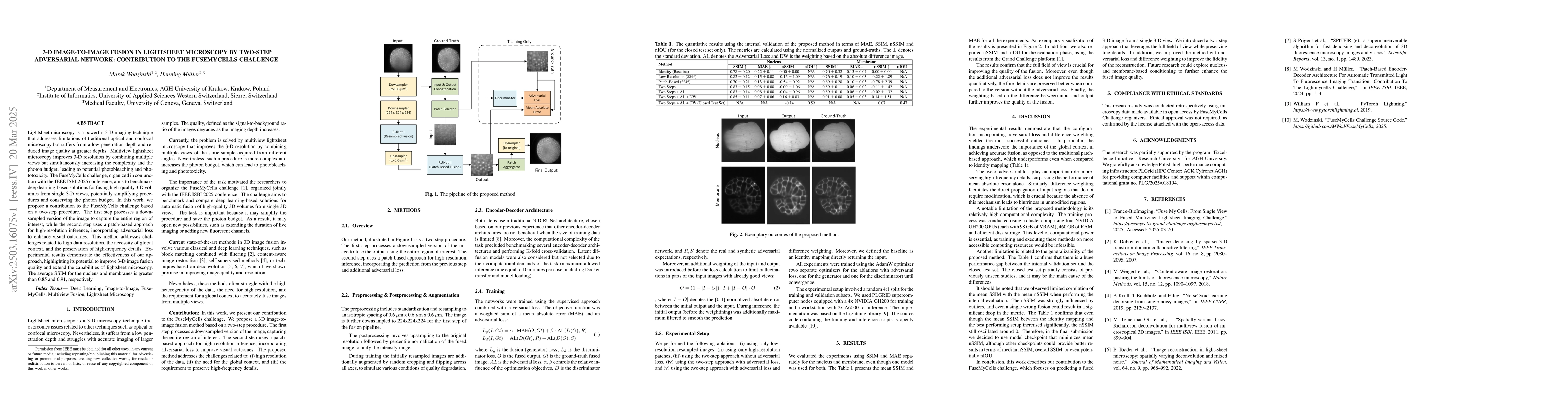
In this work, we propose a contribution to the FuseMyCells challenge based on a two-step procedure. The first step processes a downsampled version of the image to capture the entire region of interest, while the second step uses a patch-based approach for high-resolution inference, incorporating adversarial loss to enhance visual outcomes. This method addresses challenges related to high data resolution, the necessity of global context, and the preservation of high-frequency details. Experimental results demonstrate the effectiveness of our approach, highlighting its potential to improve 3-D image fusion quality and extend the capabilities of lightsheet microscopy. The average SSIM for the nucleus and membranes is greater than 0.85 and 0.91, respectively.

AI Key Findings

Generated Jun 10, 2025

Methodology

The research proposes a two-step procedure for 3-D image fusion in lightsheet microscopy, utilizing a downsampled version for global context and a patch-based approach for high-resolution inference with adversarial loss for enhanced visual outcomes.

Key Results

  • The method achieved an average SSIM greater than 0.85 for nucleus and 0.91 for membranes.
  • Experiments demonstrated the effectiveness of the approach in improving 3-D image fusion quality.
  • The two-step approach with adversarial loss and difference weighting yielded the most successful outcomes.

Significance

This research contributes to the FuseMyCells challenge, potentially simplifying lightsheet microscopy procedures and conserving the photon budget by fusing high-quality 3-D volumes from single 3-D views using deep learning.

Technical Contribution

The main technical contribution is a two-step adversarial network for 3-D image fusion in lightsheet microscopy, addressing challenges related to high data resolution, global context, and high-frequency detail preservation.

Novelty

The proposed method introduces a two-step fusion process combining downsampling for global context and patch-based high-resolution inference with adversarial loss, distinguishing it from traditional patch-based approaches and single-step methods.

Limitations

  • The method has relatively high computational complexity, requiring a cluster of four NVIDIA GH200 GPUs for training.
  • There is a significant performance gap between the internal validation set and the closed test set, indicating limited generalizability.

Future Work

  • Exploration of nucleus- and membrane-based conditioning to further enhance fused image quality.
  • Investigation into reducing computational complexity for more accessible computing resources.

Paper Details

PDF Preview

Download PDF

Key Terms

No key terms available for this paper.

No citations found for this paper.

Comments (0)

This website uses cookies to ensure you get the best experience. Learn more