Quick Summary:

This paper proposes using conditional generative adversarial networks to synthesize realistic 3D fluorescence microscopy data from cellular structure masks, enabling the creation of fully-annotated datasets for training and benchmarking deep learning models. The approach also allows for generating data of varying quality and size, demonstrating a proof-of-concept for reducing the need for extensive manual annotations.

Summary

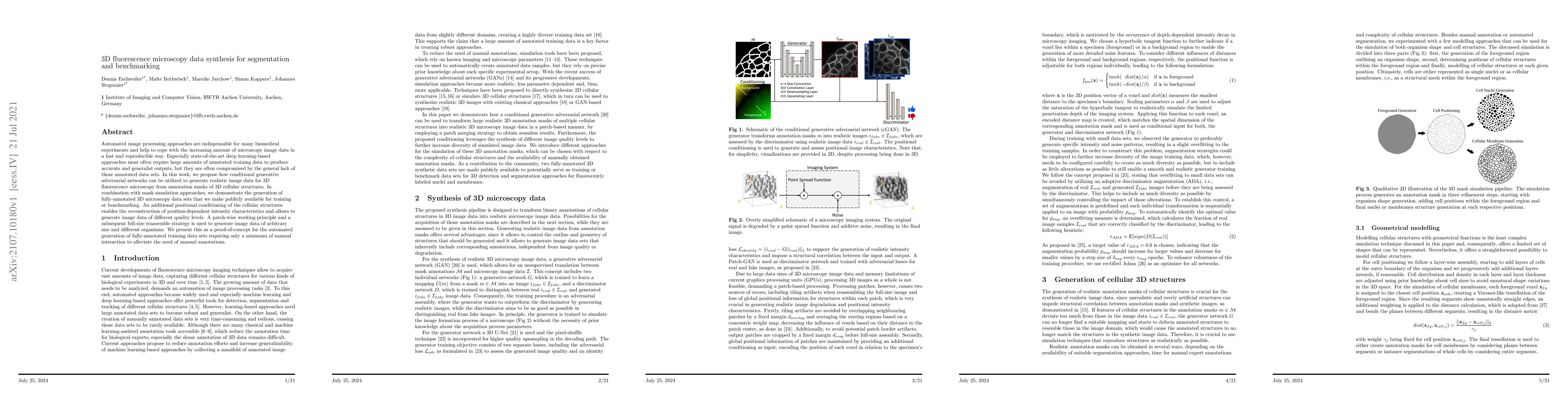
Automated image processing approaches are indispensable for many biomedical experiments and help to cope with the increasing amount of microscopy image data in a fast and reproducible way. Especially state-of-the-art deep learning-based approaches most often require large amounts of annotated training data to produce accurate and generalist outputs, but they are often compromised by the general lack of those annotated data sets. In this work, we propose how conditional generative adversarial networks can be utilized to generate realistic image data for 3D fluorescence microscopy from annotation masks of 3D cellular structures. In combination with mask simulation approaches, we demonstrate the generation of fully-annotated 3D microscopy data sets that we make publicly available for training or benchmarking. An additional positional conditioning of the cellular structures enables the reconstruction of position-dependent intensity characteristics and allows to generate image data of different quality levels. A patch-wise working principle and a subsequent full-size reassemble strategy is used to generate image data of arbitrary size and different organisms. We present this as a proof-of-concept for the automated generation of fully-annotated training data sets requiring only a minimum of manual interaction to alleviate the need of manual annotations.

AI Key Findings

Get AI-generated insights about this paper's methodology, results, and significance.

Paper Details

PDF Preview

Download PDF

Key Terms

annotated (0.268)
annotated (0.258)
microscopy (0.219)
data sets (0.216)
data sets (0.213)
microscopy (0.211)
cellular (0.192)
image (0.189)
image (0.189)
manual (0.187)
manual (0.183)
cellular (0.173)
training data (0.169)
automated (0.168)
sets (0.166)
generate (0.166)
automated (0.165)
training data (0.163)
sets (0.163)
generate (0.160)

Citation Network

Current paper (gray), citations (green), references (blue)

Current Paper
Citations
References
Click to view
Title Authors Year Actions
Cellpose as a reliable method for single-cell segmentation of autofluorescence microscopy images
Jeremiah Riendeau, A. Gillette, E. C. Guzman, M. C. Cruz, Aleksander Kralovec, Shirsa Udgata, Alexa Schmitz, Dustin A Deming, B. Cimini, Melissa C. Skala
2025
View Paper
Improving 3D deep learning segmentation with biophysically motivated cell synthesis
Roman Bruch, Mario Vitacolonna, Elina Nürnberg, Simeon Sauer, Rüdiger Rudolf, Markus Reischl
2024
View Paper
Generative adversarial networks in cell microscopy for image augmentation. A systematic review
D. Lesmes-Leon, A. Dengel, Sheraz Ahmed
2023
View Paper
AnyStar: Domain randomized universal star-convex 3D instance segmentation
Neel Dey, S. M. Abulnaga, Benjamin Billot, E. Turk, P. Grant, Adrian V. Dalca, P. Golland
2023
View Paper
Denoising diffusion probabilistic models for 3D medical image generation
Firas Khader, Gustav Mueller-Franzes, Soroosh Tayebi Arasteh, T. Han, Christoph Haarburger, M. Schulze-Hagen, P. Schad, S. Engelhardt, B. Baessler, S. Foersch, J. Stegmaier, C. Kuhl, S. Nebelung, Jakob Nikolas Kather, D. Truhn
2023
View Paper
Synthesis of large scale 3D microscopic images of 3D cell cultures for training and benchmarking
Roman Bruch, Florian Keller, Moritz Böhland, M. Vitacolonna, Lukas Klinger, R. Rudolf, M. Reischl
2023
View Paper
Inverse 3D microscopy rendering for cell shape inference with active mesh
Sacha Ichbiah, Anshuman Sinha, F. Delbary, H. Turlier
2023
View Paper
Denoising diffusion probabilistic models for generation of realistic fully-annotated microscopy image datasets
Dennis Eschweiler, J. Stegmaier
2023
View Paper
Medical Diffusion: Denoising Diffusion Probabilistic Models for 3D Medical Image Generation
Firas Khader, Gustav Mueller-Franzes, Soroosh Tayebi Arasteh, T. Han, Christoph Haarburger, M. Schulze-Hagen, P. Schad, S. Engelhardt, B. Baessler, S. Foersch, J. Stegmaier, C. Kuhl, S. Nebelung, Jakob Nikolas Kather, D. Truhn
2022
View Paper
Prediction of Fluorescent Ki67 Staining in 3D Tumor Spheroids
Roman Bruch, M. Vitacolonna, R. Rudolf, M. Reischl
2022
View Paper
Open-Source Biomedical Image Analysis Models: A Meta-Analysis and Continuous Survey
Rui Li, Vaibhav Sharma, Subasini Thangamani, A. Yakimovich
2022
View Paper
Improving Generative Adversarial Networks for Patch-Based Unpaired Image-to-Image Translation
Moritz Böhland, Roman Bruch, Simon Bäuerle, Luca Rettenberger, Markus Reischl
2023
View Paper
Medical Diffusion - Denoising Diffusion Probabilistic Models for 3D Medical Image Generation
Firas Khader, Gustav Mueller-Franzes, Soroosh Tayebi Arasteh, T. Han, Christoph Haarburger, M. Schulze-Hagen, P. Schad, Sandy Engelhardt, Bettina Baeßler, S. Foersch, Johannes Stegmaier, C. Kuhl, S. Nebelung, Jakob Nikolas Kather, D. Truhn
2022
View Paper

Comments (0)

This website uses cookies to ensure you get the best experience. Learn more