3D MRI brain tumor segmentation using autoencoder regularization
Publication
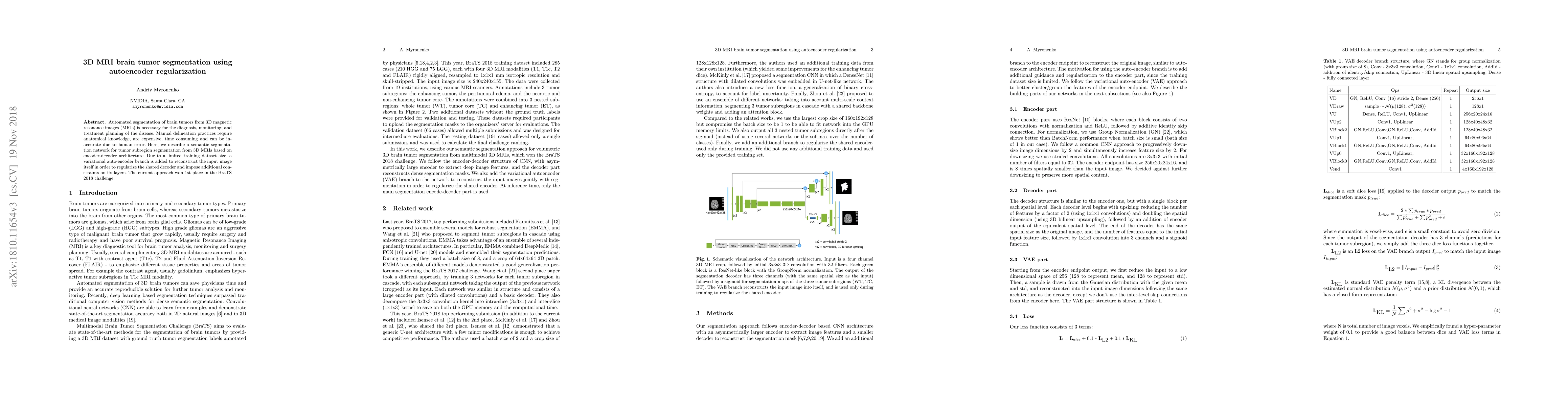
Metrics
AI Quick Summary
This paper presents a semantic segmentation network using an encoder-decoder architecture with autoencoder regularization for segmenting brain tumors in 3D MRIs. To address limited training data, a variational autoencoder reconstructs the input image, enhancing the model's performance, which won first place in the BraTS 2018 challenge.
Paper Preview
Abstract
Automated segmentation of brain tumors from 3D magnetic resonance images (MRIs) is necessary for the diagnosis, monitoring, and treatment planning of the disease. Manual delineation practices require anatomical knowledge, are expensive, time consuming and can be inaccurate due to human error. Here, we describe a semantic segmentation network for tumor subregion segmentation from 3D MRIs based on encoder-decoder architecture. Due to a limited training dataset size, a variational auto-encoder branch is added to reconstruct the input image itself in order to regularize the shared decoder and impose additional constraints on its layers. The current approach won 1st place in the BraTS 2018 challenge.
AI Key Findings
Get AI-generated insights about this paper's methodology, results, significance, and more — seven facets brought into focus.
Impact
Paper Details
PDF Preview
Key Terms
Citation Network
Current paper (gray), citations (green), references (blue)
Display is limited for performance on very large graphs.
Discussion 0