Publication
Metrics
AI Quick Summary
Researchers developed a method to automatically reconstruct 3D organ shapes from topogram images, reducing radiation exposure and improving accuracy for medical assessments.
Paper Preview
Abstract
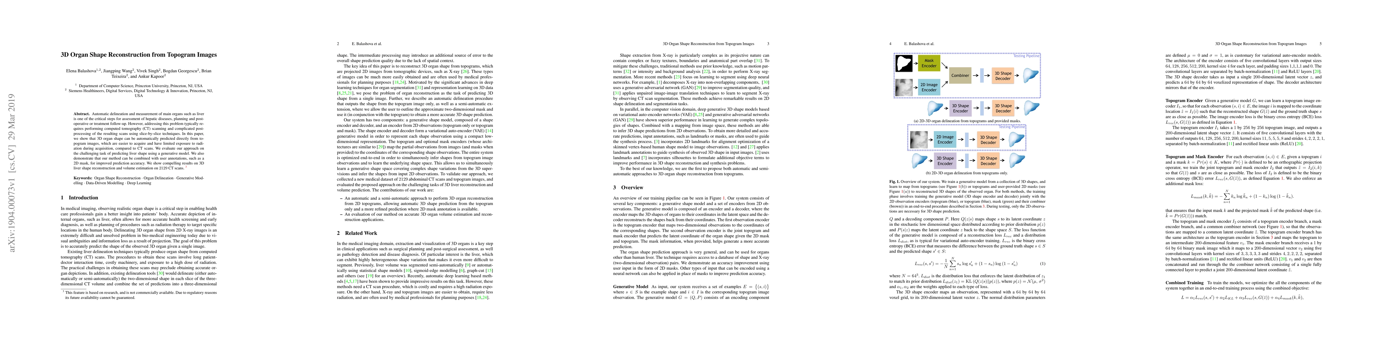
Automatic delineation and measurement of main organs such as liver is one of the critical steps for assessment of hepatic diseases, planning and postoperative or treatment follow-up. However, addressing this problem typically requires performing computed tomography (CT) scanning and complicated postprocessing of the resulting scans using slice-by-slice techniques. In this paper, we show that 3D organ shape can be automatically predicted directly from topogram images, which are easier to acquire and have limited exposure to radiation during acquisition, compared to CT scans. We evaluate our approach on the challenging task of predicting liver shape using a generative model. We also demonstrate that our method can be combined with user annotations, such as a 2D mask, for improved prediction accuracy. We show compelling results on 3D liver shape reconstruction and volume estimation on 2129 CT scans.
AI Key Findings
Get AI-generated insights about this paper's methodology, results, significance, and more — seven facets brought into focus.
Impact
Paper Details
PDF Preview
Key Terms
Citation Network
Current paper (gray), citations (green), references (blue)
Display is limited for performance on very large graphs.
Discussion 0