Summary
The purpose of this study is to present a new semi-automated methodology for three-dimensional (3D) reconstruction of coronary arteries and their plaque morphology using Computed Tomography Angiography (CTA) images. The methodology is summarized in seven stages: pre-processing of the acquired CTA images, extraction of the vessel tree centerline, estimation of a weight function for lumen, outer wall and calcified plaque, lumen segmentation, outer wall segmentation, plaque detection, and finally 3D surfaces construction. The methodology was evaluated using both expert manual annotations and estimations of a recently presented Intravascular Ultrasound (IVUS) reconstruction method. As far as the manual annotation validation process is concerned, the mean value of the comparison metrics for the 3D segmentation were 0.749 and 1.746 for the Dice coefficient and Hausdorff distance, respectively. On the other hand, the correlation coefficients for the degree of stenosis 1, the degree of stenosis 2, the plaque burden, the minimal lumen area and the minimal lumen diameter, when comparing the derived from the proposed methodology 3D models with the IVUS reconstructed models, were 0.79, 0.77, 0.75, 0.85, 0.81, respectively. The proposed methodology is an innovative approach for reconstruction of coronary arteries, since it provides 3D models of the lumen, the outer wall and the CP plaques, using the minimal user interaction. Its first implementation demonstrated that it provides an accurate reconstruction of coronary arteries and thus, it may have a wide clinical applicability
AI Key Findings
Generated Sep 02, 2025
Methodology
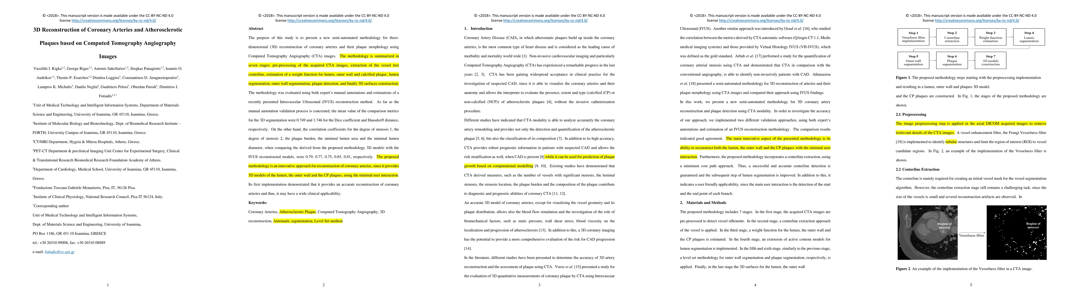
The paper presents a semi-automated methodology for 3D reconstruction of coronary arteries and atherosclerotic plaques using Computed Tomography Angiography (CTA) images, involving seven stages: pre-processing, vessel tree centerline extraction, weight function estimation, lumen segmentation, outer wall segmentation, plaque detection, and 3D surface construction.
Key Results
- The methodology was validated using expert manual annotations and Intravascular Ultrasound (IVUS) reconstructions, achieving a mean Dice coefficient of 0.749 and a mean Hausdorff distance of 1.746 with manual annotations.
- Correlation coefficients for various metrics (Degree of Stenosis 1, Degree of Stenosis 2, Plaque Burden, Minimal Lumen Area, Minimal Lumen Diameter) when comparing the proposed methodology's 3D models with IVUS reconstructions ranged from 0.75 to 0.85.
- The approach provides 3D models of the lumen, outer wall, and calcified plaques with minimal user interaction.
Significance
This research is significant as it offers a semi-automated, accurate, and detailed 3D reconstruction of coronary arteries and plaque distribution, which can aid in diagnosis and treatment planning, potentially reducing the need for invasive procedures.
Technical Contribution
The paper introduces a novel, semi-automated Level Set-based segmentation method for coronary artery reconstruction, utilizing CTA images, which provides 3D models of lumen, outer wall, and calcified plaques with minimal user input.
Novelty
This work stands out by being the first to present a 3D reconstruction approach for coronary anatomy and plaque characterization that is validated against both medical expert annotations and IVUS 3D models, offering a comprehensive evaluation of its performance.
Limitations
- The methodology does not currently account for blooming artifacts in CTA images, which can affect the accuracy of lumen contour identification.
- Parameters for discriminating lumen, outer wall, and calcified plaques are defined heuristically and could benefit from automatic adaptation to varying CTA image intensities.
Future Work
- Incorporate blooming artifact removal to enhance diagnostic confidence in vessel stenosis quantification.
- Adapt algorithm parameters automatically to different CTA image intensities for improved accuracy and user-friendliness.
Paper Details
PDF Preview
Key Terms
Citation Network
Current paper (gray), citations (green), references (blue)
Display is limited for performance on very large graphs.
Similar Papers
Found 4 papersComputed tomography coronary angiogram images, annotations and associated data of normal and diseased arteries
Arcot Sowmya, Susann Beier, Ramtin Gharleghi et al.
ImageCAS: A Large-Scale Dataset and Benchmark for Coronary Artery Segmentation based on Computed Tomography Angiography Images
Xiaomeng Li, Yiyu Shi, Xiaowei Xu et al.
| Title | Authors | Year | Actions |
|---|
Comments (0)