Summary
X-ray angiography is widely used in cardiac interventions to visualize coronary vessels, assess integrity, detect stenoses and guide treatment. We propose a framework for reconstructing 3D vessel trees from biplanar X-ray images which are extracted from two X-ray videos captured at different C-arm angles. The proposed framework consists of three main components: image segmentation, motion phase matching, and 3D reconstruction. An automatic video segmentation method for X-ray angiography to enable semantic segmentation for image segmentation and motion phase matching. The goal of the motion phase matching is to identify a pair of X-ray images that correspond to a similar respiratory and cardiac motion phase to reduce errors in 3D reconstruction. This is achieved by tracking a stationary object such as a catheter or lead within the X-ray video. The semantic segmentation approach assigns different labels to different object classes enabling accurate differentiation between blood vessels, balloons, and catheters. Once a suitable image pair is selected, key anatomical landmarks (vessel branching points and endpoints) are matched between the two views using a heuristic method that minimizes reconstruction errors. This is followed by a novel geometric reconstruction algorithm to generate the 3D vessel tree. The algorithm computes the 3D vessel centrelines by determining the intersection of two 3D surfaces. Compared to traditional methods based on epipolar constraints, the proposed approach simplifies there construction workflow and improves overall accuracy. We trained and validated our segmentation method on 62 X-ray angiography video sequences. On the test set, our method achieved a segmentation accuracy of 0.703. The 3D reconstruction framework was validated by measuring the reconstruction error of key anatomical landmarks, achieving a reprojection errors of 0.62mm +/- 0.38mm.
AI Key Findings
Generated Oct 02, 2025
Methodology
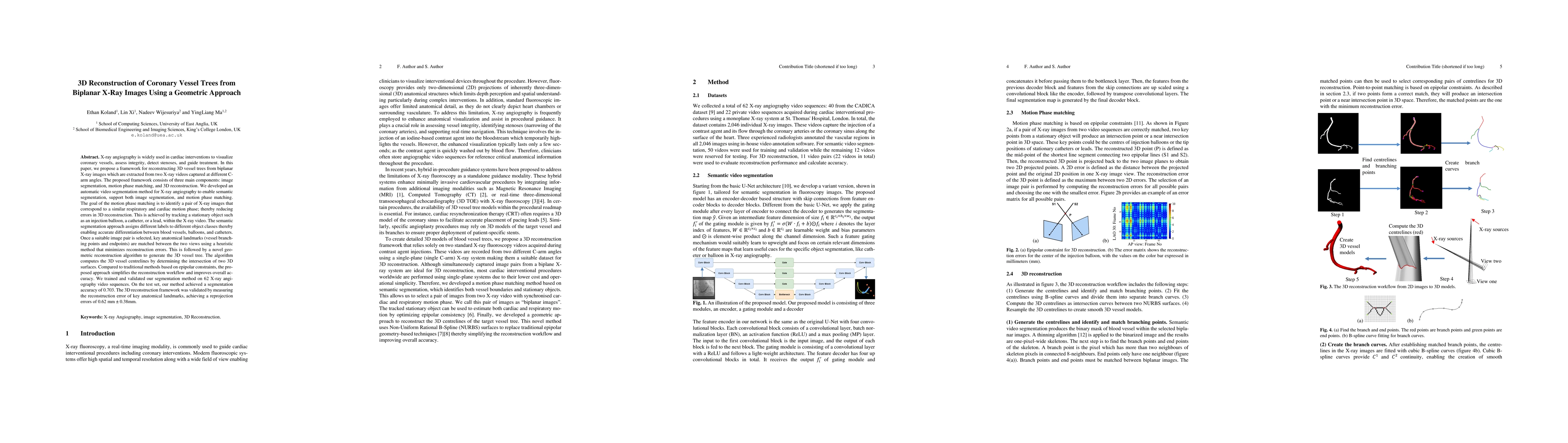
The research proposes a framework for 3D coronary vessel tree reconstruction from biplanar X-ray images using a combination of semantic video segmentation, motion phase matching, and a novel geometric reconstruction algorithm based on NURBS surfaces and epipolar constraints.
Key Results
- The segmentation method achieved a mean IoU of 0.703 on 62 X-ray angiography sequences.
- The 3D reconstruction framework achieved a reprojection error of 0.62 mm ± 0.38 mm for key anatomical landmarks.
- Motion phase matching reduced the reconstruction error of stationary object key points from 1.73 mm ± 0.96 mm to 0.18 mm ± 0.08 mm.
Significance
This research provides a faster and more accurate method for 3D coronary vessel reconstruction, which can improve cardiac interventional procedures by enabling real-time 3D guidance with reduced radiation exposure compared to traditional methods.
Technical Contribution
A novel geometric reconstruction algorithm using NURBS surfaces and intersection curves to compute 3D vessel centrelines, eliminating the need for complex post-processing steps in traditional epipolar constraint methods.
Novelty
The framework combines semantic video segmentation with motion phase matching and a geometric reconstruction approach based on NURBS surfaces, offering a streamlined workflow that improves accuracy and reduces computational complexity compared to existing methods.
Limitations
- Validation was limited to reprojection error metrics due to lack of 3D ground truth data.
- The framework was tested on a small dataset from 11 patients, limiting generalizability.
Future Work
- Integration with 3D CT scans for improved validation and ground truth comparison.
- Expansion to larger clinical datasets to improve robustness and generalization.
- Development of real-time processing capabilities for intra-procedural guidance.
Paper Details
PDF Preview
Similar Papers
Found 4 papersThree-dimensional Reconstruction of the Lumbar Spine with Submillimeter Accuracy Using Biplanar X-ray Images
Cong Wang, Yan Yu, Wanxin Yu et al.
3D Coronary Vessel Reconstruction from Bi-Plane Angiography using Graph Convolutional Networks
Qianni Zhang, Kit Mills Bransby, Greg Slabaugh et al.
Benchmarking Encoder-Decoder Architectures for Biplanar X-ray to 3D Shape Reconstruction
Bishesh Khanal, Mahesh Shakya
NeAS: 3D Reconstruction from X-ray Images using Neural Attenuation Surface
Takeshi Oishi, Chengrui Zhu, Ryoichi Ishikawa et al.
Comments (0)