Summary
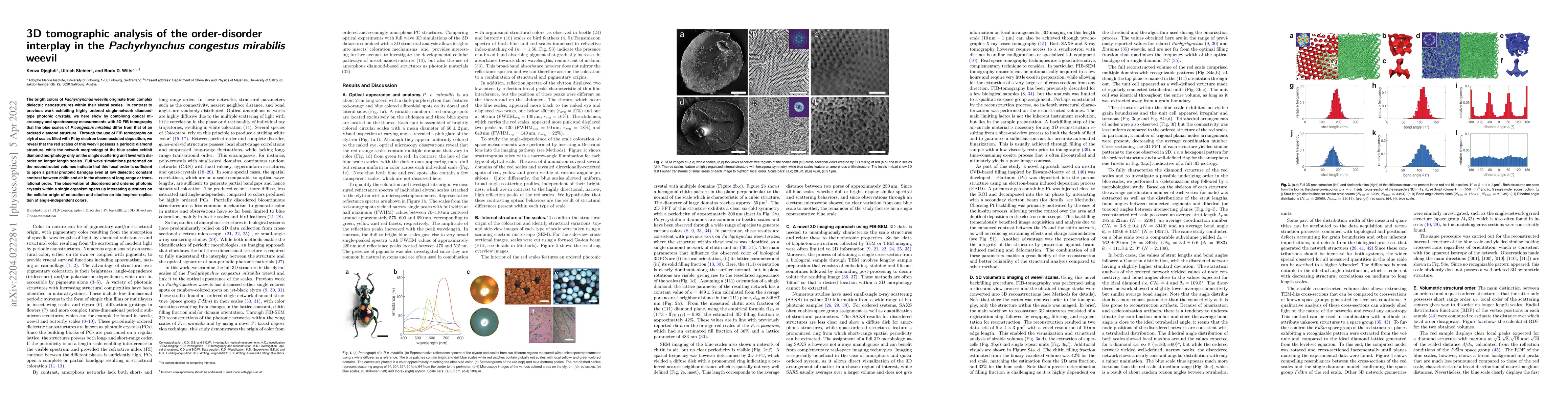
The bright colors of Pachyrhynchus weevils originate from complex dielectric nanostructures within their elytral scales. In contrast to previous work exhibiting highly ordered single-network diamond-type photonic crystals, we here show by combining optical microscopy and spectroscopy measurements with 3D FIB tomography that the blue scales of P. congestus mirabilis differ from that of an ordered diamond structure. Through the use of FIB tomography on elytral scales filled with Pt by electron beam-assisted deposition, we reveal that the red scales of this weevil possess a periodic diamond structure, while the network morphology of the blue scales exhibit diamond morphology only on the single scattering unit level with disorder on longer length scales. Full wave simulations performed on the reconstructed volumes indicate that this local order is sufficient to open a partial photonic bandgap even at low dielectric constant contrast between chitin and air in the absence of long-range or translational order. The observation of disordered and ordered photonic crystals within a single organism opens up interesting questions on the cellular origin of coloration and studies on bio-inspired replication of angle-independent colors.
AI Key Findings
Get AI-generated insights about this paper's methodology, results, and significance.
Paper Details
PDF Preview
Key Terms
Citation Network
Current paper (gray), citations (green), references (blue)
Display is limited for performance on very large graphs.
Similar Papers
Found 4 papersUsing controlled disorder to probe the interplay between charge order and superconductivity in NbSe2
Interplay of the disorder and strain in gallium oxide
Alexander Azarov, Vishnukanthan Venkatachalapathy, Andrej Kuznetsov et al.
| Title | Authors | Year | Actions |
|---|
Comments (0)