Publication
Metrics
AI Quick Summary
This study explores the feasibility of 3D ultrasound shear wave elastography for assessing musculoskeletal tissue under compressive loads, demonstrating its potential for volumetric analysis and reducing measurement bias. Preliminary results suggest it could advance computational biomechanical modeling for clinical applications.
Paper Preview
Abstract
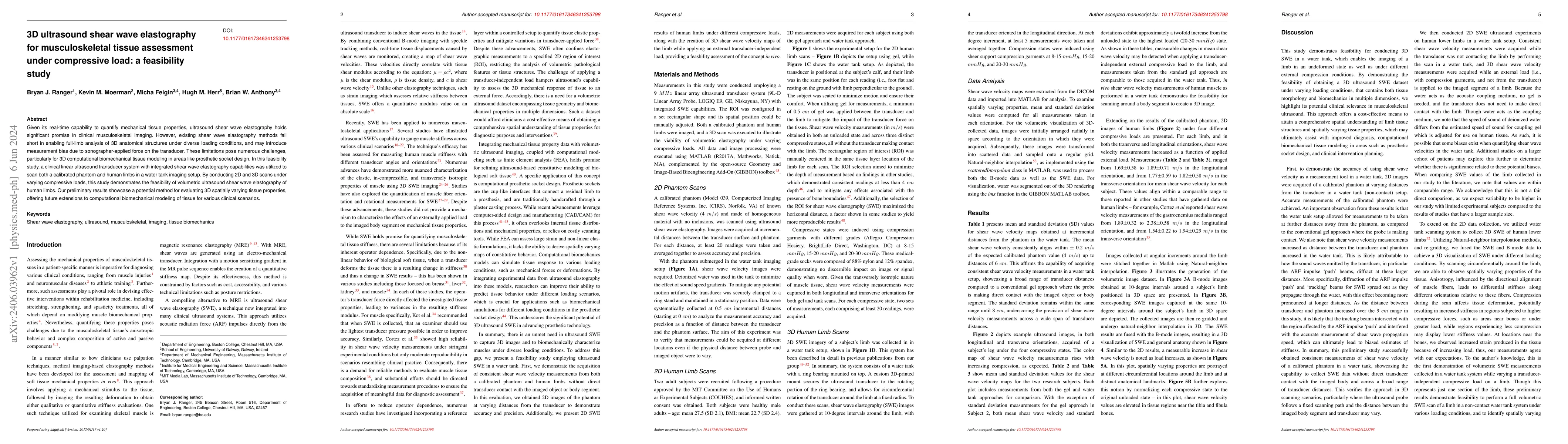
Given its real-time capability to quantify mechanical tissue properties, ultrasound shear wave elastography holds significant promise in clinical musculoskeletal imaging. However, existing shear wave elastography methods fall short in enabling full-limb analysis of 3D anatomical structures under diverse loading conditions, and may introduce measurement bias due to sonographer-applied force on the transducer. These limitations pose numerous challenges, particularly for 3D computational biomechanical tissue modeling in areas like prosthetic socket design. In this feasibility study, a clinical linear ultrasound transducer system with integrated shear wave elastography capabilities was utilized to scan both a calibrated phantom and human limbs in a water tank imaging setup. By conducting 2D and 3D scans under varying compressive loads, this study demonstrates the feasibility of volumetric ultrasound shear wave elastography of human limbs. Our preliminary results showcase a potential method for evaluating 3D spatially varying tissue properties, offering future extensions to computational biomechanical modeling of tissue for various clinical scenarios.
AI Key Findings
Get AI-generated insights about this paper's methodology, results, significance, and more — seven facets brought into focus.
Impact
Paper Details
Authors
PDF Preview
Key Terms
Citation Network
Current paper (gray), citations (green), references (blue)
Display is limited for performance on very large graphs.
Discussion 0