Summary

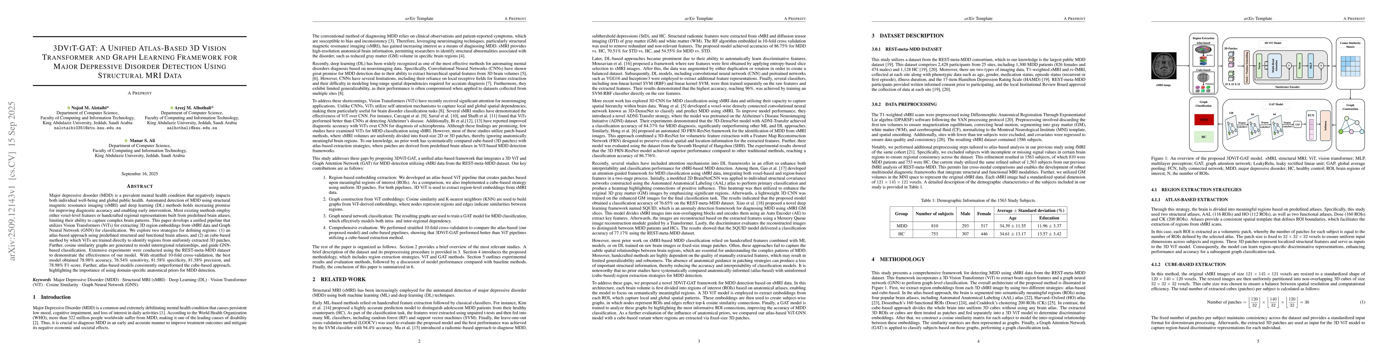
Major depressive disorder (MDD) is a prevalent mental health condition that negatively impacts both individual well-being and global public health. Automated detection of MDD using structural magnetic resonance imaging (sMRI) and deep learning (DL) methods holds increasing promise for improving diagnostic accuracy and enabling early intervention. Most existing methods employ either voxel-level features or handcrafted regional representations built from predefined brain atlases, limiting their ability to capture complex brain patterns. This paper develops a unified pipeline that utilizes Vision Transformers (ViTs) for extracting 3D region embeddings from sMRI data and Graph Neural Network (GNN) for classification. We explore two strategies for defining regions: (1) an atlas-based approach using predefined structural and functional brain atlases, and (2) an cube-based method by which ViTs are trained directly to identify regions from uniformly extracted 3D patches. Further, cosine similarity graphs are generated to model interregional relationships, and guide GNN-based classification. Extensive experiments were conducted using the REST-meta-MDD dataset to demonstrate the effectiveness of our model. With stratified 10-fold cross-validation, the best model obtained 78.98% accuracy, 76.54% sensitivity, 81.58% specificity, 81.58% precision, and 78.98% F1-score. Further, atlas-based models consistently outperformed the cube-based approach, highlighting the importance of using domain-specific anatomical priors for MDD detection.

AI Key Findings

Generated Oct 02, 2025

Methodology

The study proposes an atlas-based framework integrating 3D Vision Transformer (ViT) with Graph Attention Networks (GAT) for Major Depressive Disorder (MDD) classification using structural MRI data. The methodology involves extracting regional features using predefined brain atlases, capturing intra- and inter-regional relationships through 3D ViT, and leveraging GAT to model regional interactions for classification.

Key Results

  • Atlas-based models (AAL, HO, Dose) outperformed the cube-based model across most evaluation metrics, demonstrating the importance of anatomical priors in feature extraction.
  • The HO-based model achieved the highest accuracy (78.98%) and F1-score (78.98%) among all models, highlighting its effectiveness in capturing complex brain patterns associated with MDD.
  • Statistical comparisons showed significant differences in sensitivity, specificity, and precision between atlas-based models and the cube-based model, validating the superiority of anatomical guidance.

Significance

This research advances MDD diagnosis by leveraging anatomical priors and advanced neural network architectures to improve classification accuracy. The findings emphasize the critical role of structured, anatomically guided ROI extraction in enhancing the detection of disease-related brain changes, with potential clinical implications for early diagnosis and treatment planning.

Technical Contribution

The technical contribution lies in the integration of 3D Vision Transformer with Graph Attention Networks for the first time in MDD classification, enabling the simultaneous capture of both global and regional brain patterns while maintaining anatomical context through brain atlases.

Novelty

This work introduces a unified atlas-based framework that combines 3D ViT and GAT for MDD classification, offering a novel approach that leverages anatomical priors to enhance feature extraction and model performance compared to traditional cube-based methods.

Limitations

  • The study relies on a specific dataset (REST-Meta-MDD) which may limit generalizability to other populations.
  • The cube-based model's lack of anatomical localization may hinder its ability to detect subtle morphological patterns critical for MDD detection.

Future Work

  • Extend the framework to incorporate resting-state fMRI and clinical features for a multimodal approach.
  • Explore ensemble strategies combining information from multiple atlases to improve model generalizability.
  • Investigate the integration of additional neuroimaging modalities such as functional MRI for enhanced feature representation.

Paper Details

PDF Preview

Download PDF

Comments (0)

This website uses cookies to ensure you get the best experience. Learn more