A Bottom-Up Approach for Automatic Pancreas Segmentation in Abdominal CT Scans
Publication
Metrics
AI Quick Summary
This paper introduces a fully automated bottom-up method for pancreas segmentation in abdominal CT scans, employing a hierarchical two-tiered information propagation and a supervised random forest classifier. The method achieved 68.8% Dice coefficient and 57.2% Jaccard Index, comparable to state-of-the-art techniques.
Paper Preview
Abstract
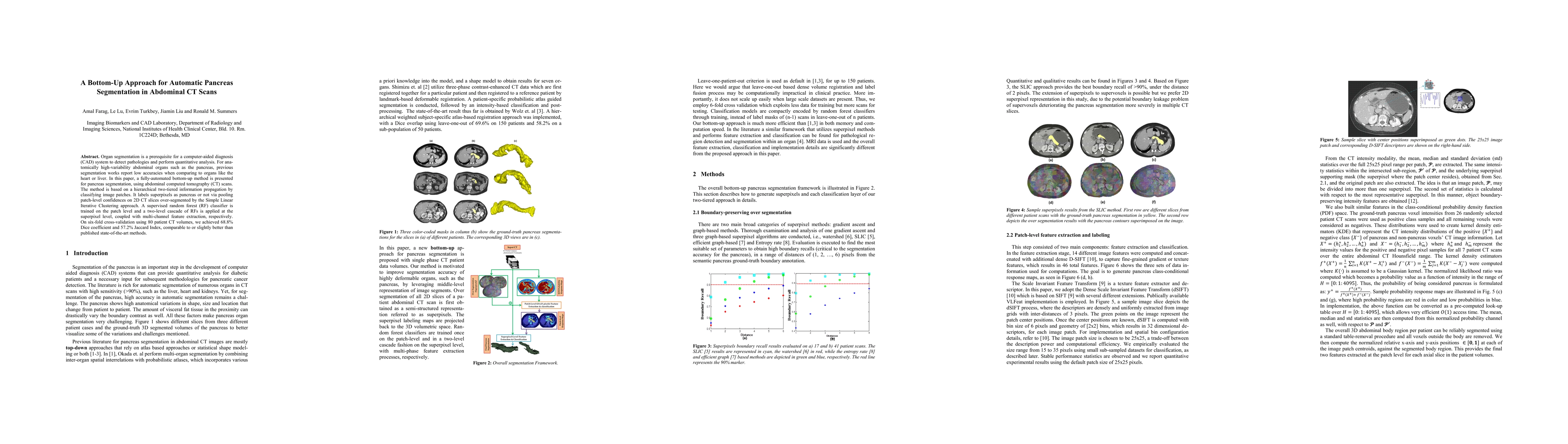
Organ segmentation is a prerequisite for a computer-aided diagnosis (CAD) system to detect pathologies and perform quantitative analysis. For anatomically high-variability abdominal organs such as the pancreas, previous segmentation works report low accuracies when comparing to organs like the heart or liver. In this paper, a fully-automated bottom-up method is presented for pancreas segmentation, using abdominal computed tomography (CT) scans. The method is based on a hierarchical two-tiered information propagation by classifying image patches. It labels superpixels as pancreas or not via pooling patch-level confidences on 2D CT slices over-segmented by the Simple Linear Iterative Clustering approach. A supervised random forest (RF) classifier is trained on the patch level and a two-level cascade of RFs is applied at the superpixel level, coupled with multi-channel feature extraction, respectively. On six-fold cross-validation using 80 patient CT volumes, we achieved 68.8% Dice coefficient and 57.2% Jaccard Index, comparable to or slightly better than published state-of-the-art methods.
AI Key Findings
Get AI-generated insights about this paper's methodology, results, significance, and more — seven facets brought into focus.
Impact
Paper Details
PDF Preview
Key Terms
Citation Network
Current paper (gray), citations (green), references (blue)
Display is limited for performance on very large graphs.
Discussion 0