A Brain Tumor Segmentation Method Based on CLIP and 3D U-Net with Cross-Modal Semantic Guidance and Multi-Level Feature Fusion
Publication
Metrics
Paper Preview
Abstract
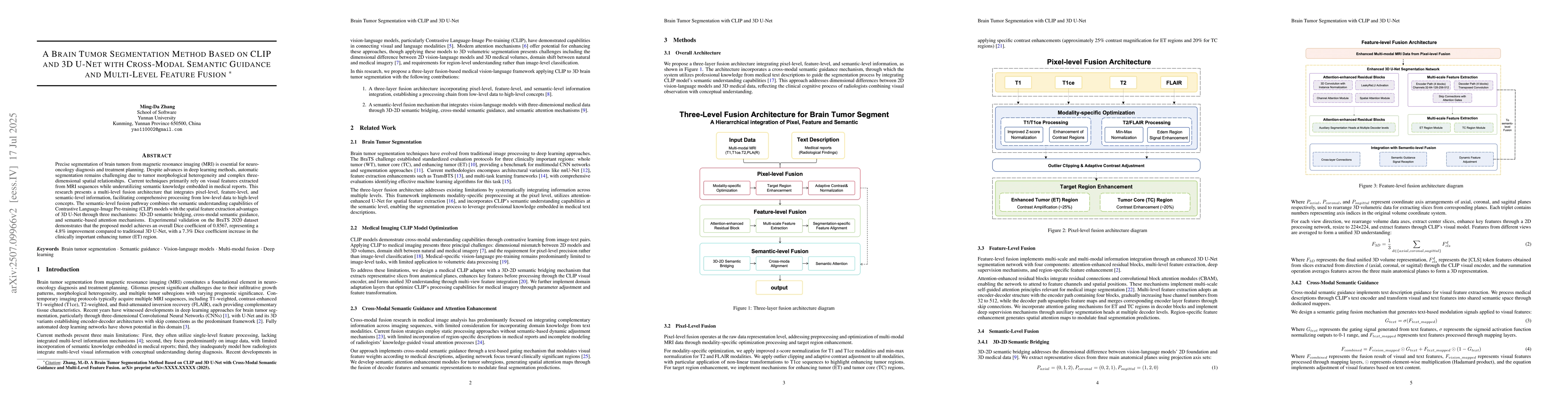
Precise segmentation of brain tumors from magnetic resonance imaging (MRI) is essential for neuro-oncology diagnosis and treatment planning. Despite advances in deep learning methods, automatic segmentation remains challenging due to tumor morphological heterogeneity and complex three-dimensional spatial relationships. Current techniques primarily rely on visual features extracted from MRI sequences while underutilizing semantic knowledge embedded in medical reports. This research presents a multi-level fusion architecture that integrates pixel-level, feature-level, and semantic-level information, facilitating comprehensive processing from low-level data to high-level concepts. The semantic-level fusion pathway combines the semantic understanding capabilities of Contrastive Language-Image Pre-training (CLIP) models with the spatial feature extraction advantages of 3D U-Net through three mechanisms: 3D-2D semantic bridging, cross-modal semantic guidance, and semantic-based attention mechanisms. Experimental validation on the BraTS 2020 dataset demonstrates that the proposed model achieves an overall Dice coefficient of 0.8567, representing a 4.8% improvement compared to traditional 3D U-Net, with a 7.3% Dice coefficient increase in the clinically important enhancing tumor (ET) region.
AI Key Findings
Get AI-generated insights about this paper's methodology, results, significance, and more — seven facets brought into focus.
Impact
Paper Details
Authors
PDF Preview
Citation Network
Current paper (gray), citations (green), references (blue)
Display is limited for performance on very large graphs.
Discussion 0