Authors
Summary
Brain tumors require an assessment to ensure timely diagnosis and effective patient treatment. Morphological factors such as size, location, texture, and variable appearance complicate tumor inspection. Medical imaging presents challenges, including noise and incomplete images. This research article presents a methodology for processing Magnetic Resonance Imaging (MRI) data, encompassing techniques for image classification and denoising. The effective use of MRI images allows medical professionals to detect brain disorders, including tumors. This research aims to categorize healthy brain tissue and brain tumors by analyzing the provided MRI data. Unlike alternative methods like Computed Tomography (CT), MRI technology offers a more detailed representation of internal anatomical components, making it a suitable option for studying data related to brain tumors. The MRI picture is first subjected to a denoising technique utilizing an Anisotropic diffusion filter. The dataset utilized for the models creation is a publicly accessible and validated Brain Tumour Classification (MRI) database, comprising 3,264 brain MRI scans. SMOTE was employed for data augmentation and dataset balancing. Convolutional Neural Networks(CNN) such as ResNet152V2, VGG, ViT, and EfficientNet were employed for the classification procedure. EfficientNet attained an accuracy of 98%, the highest recorded.
AI Key Findings
Generated Jun 11, 2025
Methodology
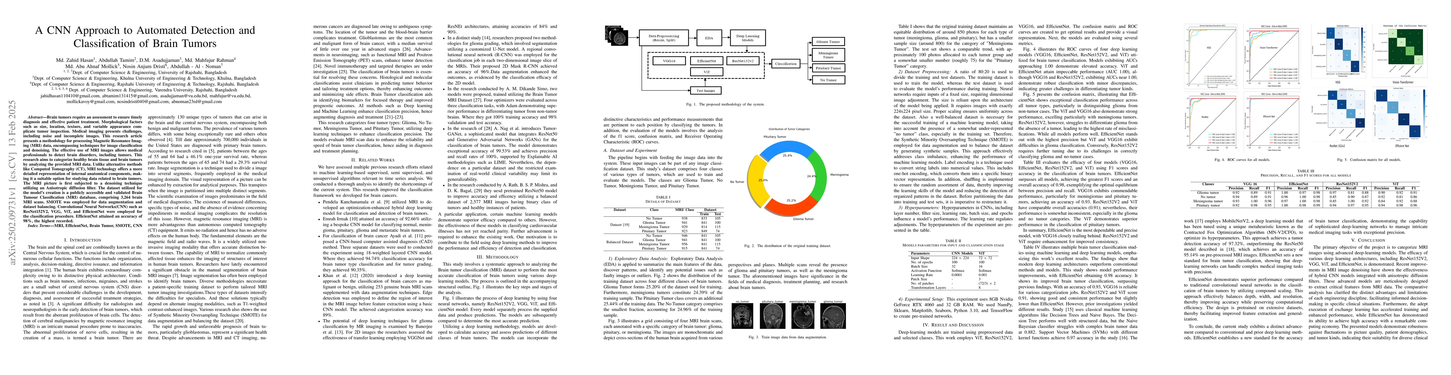
This research employs a systematic approach using deep learning models (ResNet152V2, VGG, ViT, and EfficientNet) to classify brain tumors from MRI scans. The process involves data preprocessing, including image resizing, data augmentation using SMOTE, and label encoding. Hyperparameters like epochs, batch size, and learning rate are optimized for each model.
Key Results
- EfficientNet achieved the highest accuracy of 98% in classifying brain tumors.
- ViT and EfficientNet demonstrated perfect performance (AUC 1.00) in ROC curves.
- EfficientNet showed exceptional classification performance across all tumor types, particularly distinguishing glioma from non-tumor cases.
Significance
This research contributes to the field by improving brain tumor classification accuracy using deep learning models, which can aid in diagnosis, treatment planning, and research.
Technical Contribution
The paper presents a robust deep learning methodology for brain tumor classification using various CNN architectures, showcasing EfficientNet's superior performance.
Novelty
This work distinguishes itself by employing EfficientNet, which balances depth, width, and resolution for improved accuracy and computational efficiency, outperforming traditional convolutional neural networks in brain tumor classification.
Limitations
- The study's dependence on a specific dataset may limit its generalizability.
- Real-world clinical variability was not extensively examined.
Future Work
- Further advancements are required to enhance the existing work, especially in classifying cardiovascular illnesses.
- Investigate the effectiveness of these models on other medical imaging tasks.
Paper Details
PDF Preview
Similar Papers
Found 4 papersLight Weight CNN for classification of Brain Tumors from MRI Images
Natnael Alemayehu
Detection and Classification of Brain tumors Using Deep Convolutional Neural Networks
Gopinath Balaji, Ranit Sen, Harsh Kirty
No citations found for this paper.
Comments (0)