This paper reviews computational methods for nuclei segmentation in digital pathology, tracing the evolution from traditional image processing to modern deep learning approaches. It emphasizes the importance of reducing subjectivity and time in cancer diagnosis, and discusses the potential for future research to minimize reliance on scarce annotated data while maintaining high performance.
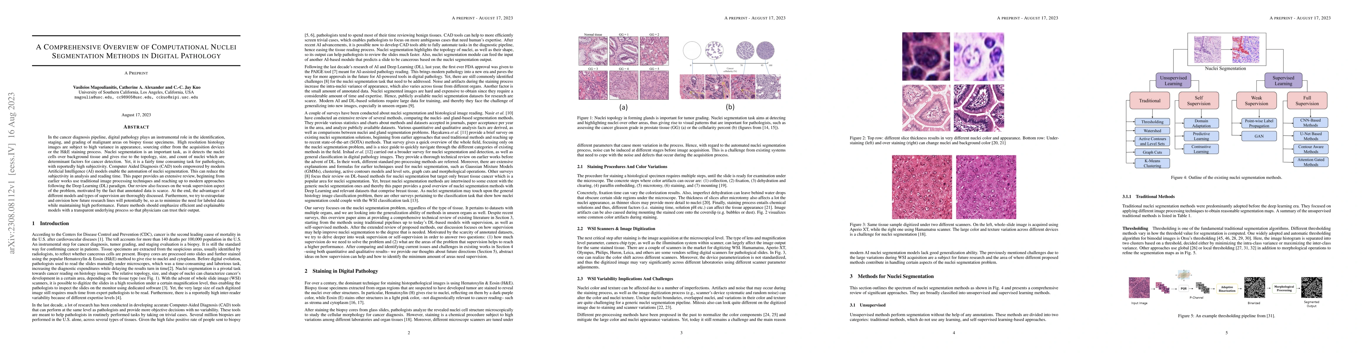
In the cancer diagnosis pipeline, digital pathology plays an instrumental role in the identification, staging, and grading of malignant areas on biopsy tissue specimens. High resolution histology images are subject to high variance in appearance, sourcing either from the acquisition devices or the H\&E staining process. Nuclei segmentation is an important task, as it detects the nuclei cells over background tissue and gives rise to the topology, size, and count of nuclei which are determinant factors for cancer detection. Yet, it is a fairly time consuming task for pathologists, with reportedly high subjectivity. Computer Aided Diagnosis (CAD) tools empowered by modern Artificial Intelligence (AI) models enable the automation of nuclei segmentation. This can reduce the subjectivity in analysis and reading time. This paper provides an extensive review, beginning from earlier works use traditional image processing techniques and reaching up to modern approaches following the Deep Learning (DL) paradigm. Our review also focuses on the weak supervision aspect of the problem, motivated by the fact that annotated data is scarce. At the end, the advantages of different models and types of supervision are thoroughly discussed. Furthermore, we try to extrapolate and envision how future research lines will potentially be, so as to minimize the need for labeled data while maintaining high performance. Future methods should emphasize efficient and explainable models with a transparent underlying process so that physicians can trust their output.
Current paper (gray), citations (green), references (blue)
Loic Le Folgoc, Pietro Gori, Julien Adam et al.
Dimitris Samaras, Alexandros Graikos, Srikar Yellapragada et al.
Ryan Erik Landvater, Ulysses Balis
Manojkumar Saranathan, Alberto Cacciola, Dianne Patterson et al.
Mohammad Sadegh Nasr, Jacob M. Luber, Jai Prakash Veerla et al.
No citations found for this paper.
Title | Authors | Year | Actions |
---|---|---|---|
Nuclei-Net: a multi-stage fusion model for nuclei segmentation in microscopy images
|
K. Roy, Subhadeep Saha, Debapriya Banik, D. Bhattacharjee
|
2023
|
View Paper |
An imbalance-aware nuclei segmentation methodology for H&E stained histopathology images
|
Emrah Hançer, Mohamed Traoré Mali, R. Samet, Z. Yildirim, Nooshin Nemati
|
2023
|
View Paper |
Impact of Stain Normalization on Pathologist Assessment of Prostate Cancer: A Comparative Study
|
M. Salvi, Alessandro Caputo, D. Balmativola, Manuela Scotto, Orazio Pennisi, Nicola Michielli, Alessandro Mogetta, F. Molinari, F. Fraggetta
|
2023
|
View Paper |
Which Pixel to Annotate: A Label-Efficient Nuclei Segmentation Framework
|
Wei Lou, Haofeng Li, Guanbin Li, Xiaoguang Han, Xiang Wan
|
2022
|
View Paper |
An Unsupervised Parameter-Free Nuclei Segmentation Method for Histology Images
|
Vasileios Magoulianitis, Peida Han, Yijing Yang, C. J. Kuo
|
2022
|
View Paper |
Nuclei and glands instance segmentation in histology images: a narrative review
|
Esha Sadia Nasir, Arshi Parvaiz, M. Fraz
|
2022
|
View Paper |
Efficient Self-Supervision using Patch-based Contrastive Learning for Histopathology Image Segmentation
|
Nicklas Boserup, Raghavendra Selvan
|
2022
|
View Paper |
A Cascaded Deep Learning Framework for Segmentation of Nuclei in Digital Histology Images
|
Khadijeh Saednia, W. Tran, A. Sadeghi-Naini
|
2022
|
View Paper |
Software tools and platforms in Digital Pathology: a review for clinicians and computer scientists
|
Rodrigo Escobar Díaz Guerrero, Lina Carvalho, T. Bocklitz, J. Popp, J. L. Oliveira
|
2022
|
View Paper |
GCP-Net: A Gating Context-Aware Pooling Network for Cervical Cell Nuclei Segmentation
|
Guihua Yang, Jinjie Huang, Yongjun He, Yuanjian Chen, Tao Wang, Cong Jin, Phienphommalinh Sengphachanh
|
2022
|
View Paper |
REU-Net: Region-enhanced nuclei segmentation network
|
Jian Qin, Yongjun He, Yang Zhou, Jing Zhao, B. Ding
|
2022
|
View Paper |
HUNIS: High-Performance Unsupervised Nuclei Instance Segmentation
|
Vasileios Magoulianitis, Yijing Yang, C.-C. Jay Kuo
|
2022
|
View Paper |
Cumulative Probability of False-Positive Results After 10 Years of Screening With Digital Breast Tomosynthesis vs Digital Mammography
|
Thao-Quyen H Ho, M. Bissell, K. Kerlikowske, R. Hubbard, B. Sprague, Christoph I. Lee, J. Tice, A. Tosteson, D. Miglioretti
|
2022
|
View Paper |
Convolutional Blur Attention Network for Cell Nuclei Segmentation
|
P. Le, Tuan D. Pham, Yi-Chiung Hsu, Jia-Ching Wang
|
2022
|
View Paper |
A region-based convolutional network for nuclei detection and segmentation in microscopy images
|
Hao-zhe Liang, Zhiming Cheng, Haiqin Zhong, Aiping Qu, Lingna Chen
|
2022
|
View Paper |
DenseRes-Unet: Segmentation of overlapped/clustered nuclei from multi organ histopathology images
|
Iqra Kiran, B. Raza, Areesha Ijaz, M. Khattak
|
2022
|
View Paper |
BT-Unet: A self-supervised learning framework for biomedical image segmentation using barlow twins with U-net models
|
N. S. Punn, Sonali Agarwal
|
2021
|
View Paper |
Mortality in the United States, 2020.
|
S. Murphy, K. Kochanek, Jiaquan Xu, E. Arias
|
2021
|
View Paper |
High-resolution deep transferred ASPPU-Net for nuclei segmentation of histopathology images
|
Amit Kumar Chanchal, S. Lal, Jyoti Kini
|
2021
|
View Paper |
Multi-channel Chan-Vese model for unsupervised segmentation of nuclei from breast histopathological images
|
R. Rashmi, K. Prasad, C. B. Udupa
|
2021
|
View Paper |
A review and comparison of breast tumor cell nuclei segmentation performances using deep convolutional neural networks
|
A. Lagree, M. Mohebpour, Nicholas Meti, Khadijeh Saednia, F. Lu, E. Slodkowska, S. Gandhi, E. Rakovitch, A. Shenfield, A. Sadeghi-Naini, W. Tran
|
2021
|
View Paper |
DARCNN: Domain Adaptive Region-based Convolutional Neural Network for Unsupervised Instance Segmentation in Biomedical Images
|
Joy Hsu, Wah Chiu, Serena Yeung
|
2021
|
View Paper |
Quantifying the Scanner-Induced Domain Gap in Mitosis Detection
|
M. Aubreville, C. Bertram, M. Veta, R. Klopfleisch, N. Stathonikos, Katharina Breininger, N. T. Hoeve, F. Ciompi, A. Maier
|
2021
|
View Paper |
A new complete color normalization method for H&E stained histopatholgical images
|
Surbhi Vijh, M. Saraswat, Sumit Kumar
|
2021
|
View Paper |
CPP-Net: Context-Aware Polygon Proposal Network for Nucleus Segmentation
|
Shengcong Chen, Changxing Ding, Minfeng Liu, Jun Cheng, D. Tao
|
2021
|
View Paper |
NucleiSegNet: Robust deep learning architecture for the nuclei segmentation of liver cancer histopathology images
|
S. Lal, Devikalyan Das, Kumar Alabhya, A. Kanfade, Aman Kumar, J. Kini
|
2020
|
View Paper |
Generative Adversarial Training for Weakly Supervised Nuclei Instance Segmentation
|
Wei Hu, Huanhuan Sheng, Jing Wu, Yining Li, Tianyi Liu, Yonghao Wang, Yuan Wen
|
2020
|
View Paper |
Weakly-Supervised Nucleus Segmentation Based on Point Annotations: A Coarse-to-Fine Self-Stimulated Learning Strategy
|
Kuan Tian, Jun Zhang, Haocheng Shen, Kezhou Yan, Pei Dong, Jianhua Yao, Shannon Che, P. Luo, Xiao Han
|
2020
|
View Paper |
Instance-aware Self-supervised Learning for Nuclei Segmentation
|
Xinpeng Xie, Jiawei Chen, Yuexiang Li, Linlin Shen, Kai Ma, Yefeng Zheng
|
2020
|
View Paper |
Self-Supervised Nuclei Segmentation in Histopathological Images Using Attention
|
M. Sahasrabudhe, S. Christodoulidis, R. Salgado, S. Michiels, S. Loi, Fabrice Andr'e, N. Paragios, M. Vakalopoulou
|
2020
|
View Paper |
Weakly Supervised Deep Nuclei Segmentation Using Partial Points Annotation in Histopathology Images
|
Hui Qu, Pengxiang Wu, Qiaoying Huang, Jingru Yi, Zhennan Yan, Kang Li, G. Riedlinger, S. De, Shaoting Zhang, Dimitris N. Metaxas
|
2020
|
View Paper |
Boundary-assisted Region Proposal Networks for Nucleus Segmentation
|
Shengcong Chen, Changxing Ding, D. Tao
|
2020
|
View Paper |
Independent validation of paige prostate: Assessing clinical benefit of an artificial intelligence tool within a digital diagnostic pathology laboratory workflow.
|
Christopher Kanan, J. Sue, L. Grady, Thomas J. Fuchs, S. Chandarlapaty, J. Reis-Filho, P. Salles, L. D. da Silva, C. Ferreira, E. Pereira
|
2020
|
View Paper |
Unsupervised Instance Segmentation in Microscopy Images via Panoptic Domain Adaptation and Task Re-Weighting
|
Dongnan Liu, Donghao Zhang, Yang Song, Fan Zhang, L. O’Donnell, Heng Huang, Mei Chen, Weidong (Tom) Cai
|
2020
|
View Paper |
A Multi-Organ Nucleus Segmentation Challenge
|
Neeraj Kumar, R. Verma, Deepak Anand, Yanning Zhou, O. F. Onder, E. Tsougenis, Hao Chen, P. Heng, Jiahui Li, Zhiqiang Hu, Yunzhi Wang, Navid Alemi Koohbanani, M. Jahanifar, Neda Zamani Tajeddin, A. Gooya, N. Rajpoot, Xuhua Ren, Sihang Zhou, Qian Wang, D. Shen, Cheng-Kun Yang, C. Weng, Wei-Hsiang Yu, Chao-Yuan Yeh, Shuang Yang, Shuoyu Xu, P. Yeung, Peng Sun, A. Mahbod, G. Schaefer, I. Ellinger, R. Ecker, O. Smedby, Chunliang Wang, Benjamin Chidester, That-Vinh Ton, M. Tran, Jian Ma, M. Do, S. Graham, Q. Vu, J. T. Kwak, Akshaykumar Gunda, R. Chunduri, Corey Hu, Xiao-xiao Zhou, Dariush Lotfi, Reza Safdari, Antanas Kascenas, Alison Q. O'Neil, Dennis Eschweiler, J. Stegmaier, Yanping Cui, Baocai Yin, Kailin Chen, Xinmei Tian, P. Gruening, E. Barth, E. Arbel, I. Remer, A. Ben-Dor, E. Sirazitdinova, Matthias Kohl, S. Braunewell, Yuexiang Li, Xinpeng Xie, Linlin Shen, Jun Ma, K. Baksi, Mohammad Azam Khan, J. Choo, Adrián Colomer, V. Naranjo, L. Pei, K. Iftekharuddin, K. Roy, D. Bhattacharjee, Aníbal Pedraza, M. G. Bueno, S. Devanathan, Saravanan Radhakrishnan, Praveen Koduganty, Zihan Wu, Guanyu Cai, Xiaojie Liu, Yuqin Wang, A. Sethi
|
2020
|
View Paper |
Nuclei Segmentation Using Mixed Points and Masks Selected From Uncertainty
|
Hui Qu, Jingru Yi, Qiaoying Huang, Pengxiang Wu, Dimitris N. Metaxas
|
2020
|
View Paper |
A Comprehensive Review for Breast Histopathology Image Analysis Using Classical and Deep Neural Networks
|
Xiaomin Zhou, Chen Li, M. Rahaman, Yudong Yao, Shiliang Ai, Changhao Sun, Qian Wang, Yong Zhang, Mo Li, Xiaoyan Li, Tao Jiang, Dan Xue, Shouliang Qi, Yueyang Teng
|
2020
|
View Paper |
A large-scale dataset for mitotic figure assessment on whole slide images of canine cutaneous mast cell tumor
|
C. Bertram, M. Aubreville, C. Marzahl, A. Maier, R. Klopfleisch
|
2019
|
View Paper |
Nucleus segmentation across imaging experiments: the 2018 Data Science Bowl
|
Juan C. Caicedo, A. Goodman, Kyle W. Karhohs, B. Cimini, Jeanelle Ackerman, Marzieh Haghighi, Cherkeng Heng, Tim Becker, M. Doan, C. McQuin, M. Rohban, Shantanu Singh, Anne E Carpenter
|
2019
|
View Paper |
Computational Nuclei Segmentation Methods in Digital Pathology: A Survey
|
Tomohiro Hayakawa, V. Prasath, H. Kawanaka, Bruce J. Aronow, Bruce J. Aronow, S. Tsuruoka
|
2019
|
View Paper |
Direct Cellularity Estimation on Breast Cancer Histopathology Images Using Transfer Learning
|
Ziang Pei, S. Cao, Lijun Lu, Wufan Chen
|
2019
|
View Paper |
PseudoEdgeNet: Nuclei Segmentation only with Point Annotations
|
Inwan Yoo, Donggeun Yoo, K. Paeng
|
2019
|
View Paper |
Weakly Supervised Deep Nuclei Segmentation using Points Annotation in Histopathology Images
|
Hui Qu, Pengxiang Wu, Qiaoying Huang, Jingru Yi, G. Riedlinger, S. De, Dimitris N. Metaxas
|
2019
|
View Paper |
Diversify and Match: A Domain Adaptive Representation Learning Paradigm for Object Detection
|
Taekyung Kim, Minki Jeong, Seunghyeon Kim, Seokeon Choi, Changick Kim
|
2019
|
View Paper |
Computational normalization of H&E-stained histological images: Progress, challenges and future potential
|
T. A. Tosta, P. D. Faria, L. A. Neves, M. Z. Nascimento
|
2019
|
View Paper |
CIA-Net: Robust Nuclei Instance Segmentation with Contour-aware Information Aggregation
|
Yanning Zhou, O. F. Onder, Q. Dou, E. Tsougenis, Hao Chen, P. Heng
|
2019
|
View Paper |
False positives in PIRADS (V2) 3, 4, and 5 lesions: relationship with reader experience and zonal location
|
T. T. Stolk, I. D. de Jong, T. Kwee, H. Luiting, S. Mahesh, B. Doornweerd, P. Willemse, Derya Yakar
|
2019
|
View Paper |
Segmentation of Nuclei in Histopathology Images by Deep Regression of the Distance Map
|
Peter Naylor, M. Laé, F. Reyal, Thomas Walter
|
2019
|
View Paper |
Hover-Net: Simultaneous segmentation and classification of nuclei in multi-tissue histology images
|
S. Graham, Q. Vu, S. Raza, A. Azam, Y. Tsang, J. T. Kwak, N. Rajpoot
|
2018
|
View Paper |
A study about color normalization methods for histopathology images.
|
Santanu Roy, Alok Kumar Jain, S. Lal, J. Kini
|
2018
|
View Paper |
Methods for Segmentation and Classification of Digital Microscopy Tissue Images
|
Q. Vu, S. Graham, Minh Nguyen Nhat To, M. Shaban, Talha Qaiser, Navid Alemi Koohbanani, S. Khurram, T. Kurç, K. Farahani, Tianhao Zhao, Rajarsi R. Gupta, J. T. Kwak, N. Rajpoot, J. Saltz
|
2018
|
View Paper |
BESNet: Boundary-Enhanced Segmentation of Cells in Histopathological Images
|
H. Oda, H. Roth, Kosuke Chiba, Jure Sokolić, T. Kitasaka, M. Oda, Akinari Hinoki, H. Uchida, J. Schnabel, K. Mori
|
2018
|
View Paper |
Attention Gated Networks: Learning to Leverage Salient Regions in Medical Images
|
Jo Schlemper, O. Oktay, M. Schaap, M. Heinrich, Bernhard Kainz, Ben Glocker, D. Rueckert
|
2018
|
View Paper |
Fast and robust segmentation of white blood cell images by self-supervised learning.
|
Xin Zheng, Yong Wang, Guoyou Wang, Jianguo Liu
|
2018
|
View Paper |
Path Aggregation Network for Instance Segmentation
|
Shu Liu, Lu Qi, Haifang Qin, Jianping Shi, Jiaya Jia
|
2018
|
View Paper |
Panoptic Segmentation
|
Alexander Kirillov, Kaiming He, Ross B. Girshick, C. Rother, Piotr Dollár
|
2018
|
View Paper |
K mean clustering based automated segmentation of overlapping cell nuclei in pleural effusion cytology images
|
Khin Yadanar Win, S. Choomchuay, K. Hamamoto
|
2017
|
View Paper |
Stain Normalization using Sparse AutoEncoders (StaNoSA): Application to digital pathology
|
A. Janowczyk, A. Basavanhally, A. Madabhushi
|
2017
|
View Paper |
Mask R-CNN
|
Kaiming He, Georgia Gkioxari, Piotr Dollár, Ross B. Girshick
|
2017
|
View Paper |
A Dataset and a Technique for Generalized Nuclear Segmentation for Computational Pathology
|
Neeraj Kumar, R. Verma, Sanuj Sharma, S. Bhargava, Abhishek Vahadane, A. Sethi
|
2017
|
View Paper |
Automated segmentation of cell nuclei in cytology pleural fluid images using OTSU thresholding
|
Khin Yadanar Win, S. Choomchuay
|
2017
|
View Paper |
DCAN: Deep contour‐aware networks for object instance segmentation from histology images
|
Hao Chen, Xiaojuan Qi, Lequan Yu, Q. Dou, J. Qin, P. Heng
|
2017
|
View Paper |
Self-adjusting nuclei segmentation (SANS) of Hematoxylin-Eosin stained histopathological breast cancer images
|
Yuxin Cui, Jianjun Hu
|
2016
|
View Paper |
An automatic system for cell nuclei pleomorphism segmentation in histopathological images of breast cancer
|
Pegah Faridi, H. Danyali, M. Helfroush, M. A. Jahromi
|
2016
|
View Paper |
Spatial Shape Constrained Fuzzy C-Means (FCM) Clustering for Nucleus Segmentation in Pap Smear Images
|
Ratna Saha, M. Bajger, Gobert N. Lee
|
2016
|
View Paper |
Automatic classification of leukocytes using morphological features and Naïve Bayes classifier
|
Anjali Gautam, Priyanka Singh, B. Raman, H. Bhadauria
|
2016
|
View Paper |
Densely Connected Convolutional Networks
|
Gao Huang, Zhuang Liu, Kilian Q. Weinberger
|
2016
|
View Paper |
Quantitative analysis of histological tissue image based on cytological profiles and spatial statistics
|
Y. Chang, Guillaume Thibault, Vahid Azimi, Brett E. Johnson, D. Jorgens, Jason M. Link, Adam A. Margolin, J. Gray
|
2016
|
View Paper |
Variability in Pathologists' Interpretations of Individual Breast Biopsy Slides: A Population Perspective
|
J. Elmore, H. Nelson, M. Pepe, G. Longton, A. Tosteson, B. Geller, T. Onega, P. Carney, Sara L. Jackson, K. Allison, D. Weaver
|
2016
|
View Paper |
Nucleus segmentation in histology images with hierarchical multilevel thresholding
|
H. A. Phoulady, Dmitry Goldgof, L. Hall, P. Mouton
|
2016
|
View Paper |
Iterative h‐minima‐based marker‐controlled watershed for cell nucleus segmentation
|
C. Koyuncu, Ece Akhan, Tulin Ersahin, R. Cetin-Atalay, C. Gunduz-Demir
|
2016
|
View Paper |
Locality Sensitive Deep Learning for Detection and Classification of Nuclei in Routine Colon Cancer Histology Images
|
K. Sirinukunwattana, S. Raza, Y. Tsang, D. Snead, I. Cree, N. Rajpoot
|
2016
|
View Paper |
An Automatic Learning-Based Framework for Robust Nucleus Segmentation
|
Fuyong Xing, Yuanpu Xie, L. Yang
|
2016
|
View Paper |
Deep Residual Learning for Image Recognition
|
Kaiming He, X. Zhang, Shaoqing Ren, Jian Sun
|
2015
|
View Paper |
Nucleus and cytoplasm segmentation in microscopic images using K-means clustering and region growing
|
Omid Sarrafzadeh, A. M. Dehnavi
|
2015
|
View Paper |
U-Net: Convolutional Networks for Biomedical Image Segmentation
|
O. Ronneberger, P. Fischer, T. Brox
|
2015
|
View Paper |
Very Deep Convolutional Networks for Large-Scale Image Recognition
|
K. Simonyan, Andrew Zisserman
|
2014
|
View Paper |
Segmentation of cytoplasm and nuclei of abnormal cells in cervical cytology using global and local graph cuts
|
Ling Zhang, Hui Kong, C. Chin, Shaoxiong Liu, Zhi Chen, Tianfu Wang, Siping Chen
|
2014
|
View Paper |
Detection of centroblasts in H&E stained images of follicular lymphoma
|
E. Michail, E. Kornaropoulos, K. Dimitropoulos, N. Grammalidis, T. Koletsa, I. Kostopoulos
|
2014
|
View Paper |
A New Iterative Triclass Thresholding Technique in Image Segmentation
|
Hongmin Cai, Zhong Yang, Xinhua Cao, W. Xia, Xiaoyin Xu
|
2014
|
View Paper |
The Wonderful Colors of the Hematoxylin–Eosin Stain in Diagnostic Surgical Pathology
|
J. Chan
|
2014
|
View Paper |
Towards generalized nuclear segmentation in histological images
|
Abhishek Vahadane, A. Sethi
|
2013
|
View Paper |
Segmenting overlapping cell nuclei in digital histopathology images
|
Jie Shu, Hao Fu, G. Qiu, P. Kaye, M. Ilyas
|
2013
|
View Paper |
A robust automatic nuclei segmentation technique for quantitative histopathological image analysis.
|
Cheng Lu, Muhammad Mahmood, N. Jha, M. Mandal
|
2012
|
View Paper |
Fiji: an open-source platform for biological-image analysis
|
J. Schindelin, Ignacio Arganda-Carreras, E. Frise, V. Kaynig, M. Longair, T. Pietzsch, S. Preibisch, C. Rueden, S. Saalfeld, Benjamin Schmid, J. Tinevez, D. J. White, V. Hartenstein, K. Eliceiri, P. Tomančák, Albert Cardona
|
2012
|
View Paper |
Marker-controlled watershed segmentation of nuclei in H&E stained breast cancer biopsy images
|
M. Veta, A. Huisman, M. Viergever, P. Diest, J. Pluim
|
2011
|
View Paper |
Improved Automatic Detection and Segmentation of Cell Nuclei in Histopathology Images
|
Yousef Al-Kofahi, W. Lassoued, William Lee, B. Roysam
|
2010
|
View Paper |
Expectation–Maximization-Driven Geodesic Active Contour With Overlap Resolution (EMaGACOR): Application to Lymphocyte Segmentation on Breast Cancer Histopathology
|
Hussain Fatakdawala, Jun Xu, A. Basavanhally, G. Bhanot, S. Ganesan, M. Feldman, John Tomaszewski, A. Madabhushi
|
2010
|
View Paper |
Histopathological Image Analysis: A Review
|
M. Gurcan, L. Boucheron, A. Can, A. Madabhushi, N. Rajpoot, B. Yener
|
2009
|
View Paper |
Segmentation of Touching Cell Nuclei Using a Two-Stage Graph Cut Model
|
O. Daněk, P. Matula, C. Ortíz-de-Solórzano, A. Muñoz-Barrutia, Martin Maška, M. Kozubek
|
2009
|
View Paper |
Hematoxylin and eosin staining of tissue and cell sections.
|
A. Fischer, K. Jacobson, Jack Rose, R. Zeller
|
2008
|
View Paper |
CellProfiler: image analysis software for identifying and quantifying cell phenotypes
|
Anne E Carpenter, T. Jones, M. R. Lamprecht, C. Clarke, In Han Kang, O. Friman, D. Guertin, Joo Han Chang, Robert A Lindquist, J. Moffat, P. Golland, D. Sabatini
|
2006
|
View Paper |
Automated cell nucleus segmentation using improved snake
|
Min Hu, X. Ping, Yihong Ding
|
2004
|
View Paper |
Active contours without edges
|
T. Chan, L. Vese
|
2001
|
View Paper |
Adaptive document image binarization
|
J. Sauvola, M. Pietikäinen
|
2000
|
View Paper |
Optimal approximations by piecewise smooth functions and associated variational problems
|
D. Mumford, J. Shah
|
1989
|
View Paper |
A Multi-scale Self-supervision Method for Improving Cell Nuclei Segmentation in Pathological Tissues
|
Hesham F. Ali, M. A. Alattar, Sahar Selim
|
2022
|
View Paper |
Label Propagation for Annotation-Efficient Nuclei Segmentation from Pathology Images
|
Yi-Mou Lin, Z. Qu, Hao Chen, Zhongke Gao, Yuexiang Li, Lili Xia, Kai Ma, Yefeng Zheng, Kwang-Ting Cheng
|
2022
|
View Paper |
Supervised Classification of Breast Cancer Malignancy Using Integrated Modified Marker Controlled Watershed Approach
|
Rajyalakshmi Uppada, S. Rao, K. Prasad
|
2017
|
View Paper |
Automatic segmentation of cell nuclei using Krill Herd optimization based multi-thresholding and Localized Active Contour Model
|
S. Beevi K, Madhu S. Nair, G. R. Bindu
|
2016
|
View Paper |
Methods for Nuclei Detection, Segmentation, and Classification in Digital Histopathology: A Review—Current Status and Future Potential
|
H. Irshad, A. Veillard, L. Roux, Daniel Racoceanu
|
2014
|
View Paper |
“Tratado de histologia em cores,”
|
2007
|
||
Image Analysis for Neuroblastoma Classification: Segmentation of Cell Nuclei
|
M. Gurcan, T. Pan, H. Shimada, J. Saltz
|
2006
|
View Paper |
The Watershed Transform: Definitions, Algorithms and Parallelization Strategies
|
2000
|
View Paper | |
A Threshold Selection Method from Gray-Level Histograms
|
N. Otsu
|
1979
|
View Paper |
Showing first page preview. Click "Open Full PDF" to view the complete paper.
Your comment contains inappropriate language and has been rejected.
Are you sure you want to remove this tag? This action cannot be undone.
Someone just favorited a paper!
Comments (0)