A Context-aware Delayed Agglomeration Framework for Electron Microscopy Segmentation
Publication
Metrics
AI Quick Summary
A new framework for electron microscopy image segmentation outperforms existing methods with improved accuracy on 2D and 3D datasets.
Paper Preview
Abstract
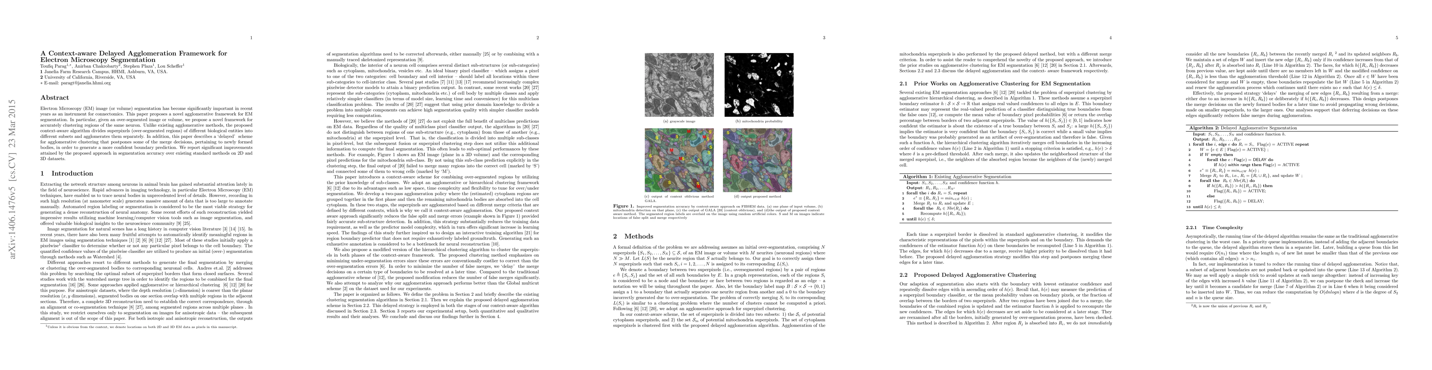
Electron Microscopy (EM) image (or volume) segmentation has become significantly important in recent years as an instrument for connectomics. This paper proposes a novel agglomerative framework for EM segmentation. In particular, given an over-segmented image or volume, we propose a novel framework for accurately clustering regions of the same neuron. Unlike existing agglomerative methods, the proposed context-aware algorithm divides superpixels (over-segmented regions) of different biological entities into different subsets and agglomerates them separately. In addition, this paper describes a "delayed" scheme for agglomerative clustering that postpones some of the merge decisions, pertaining to newly formed bodies, in order to generate a more confident boundary prediction. We report significant improvements attained by the proposed approach in segmentation accuracy over existing standard methods on 2D and 3D datasets.
AI Key Findings
Get AI-generated insights about this paper's methodology, results, significance, and more — seven facets brought into focus.
Impact
Paper Details
PDF Preview
Key Terms
Citation Network
Current paper (gray), citations (green), references (blue)
Display is limited for performance on very large graphs.
Discussion 0