A Contrast Synthesized Thalamic Nuclei Segmentation Scheme using Convolutional Neural Networks
Publication
Metrics
AI Quick Summary
This paper explores 3D Convolutional Neural Networks (CNNs) for segmenting thalamic nuclei from conventional MPRAGE images, comparing native contrast segmentation (NCS) with synthesized contrast segmentation (SCS) using synthesized WMn-MPRAGE images. The SCS network showed higher accuracy and clinical relevance, particularly in detecting atrophy in alcohol use disorder patients.
Paper Preview
Abstract
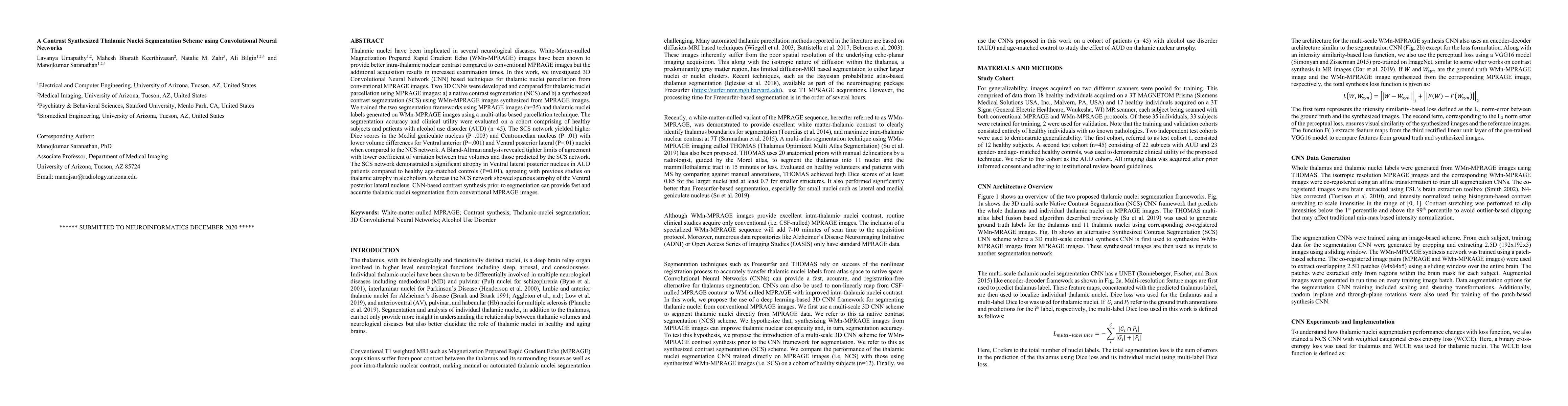
Thalamic nuclei have been implicated in several neurological diseases. WMn-MPRAGE images have been shown to provide better intra-thalamic nuclear contrast compared to conventional MPRAGE images but the additional acquisition results in increased examination times. In this work, we investigated 3D Convolutional Neural Network (CNN) based techniques for thalamic nuclei parcellation from conventional MPRAGE images. Two 3D CNNs were developed and compared for thalamic nuclei parcellation using MPRAGE images: a) a native contrast segmentation (NCS) and b) a synthesized contrast segmentation (SCS) using WMn-MPRAGE images synthesized from MPRAGE images. We trained the two segmentation frameworks using MPRAGE images (n=35) and thalamic nuclei labels generated on WMn-MPRAGE images using a multi-atlas based parcellation technique. The segmentation accuracy and clinical utility were evaluated on a cohort comprising of healthy subjects and patients with alcohol use disorder (AUD) (n=45). The SCS network yielded higher Dice scores in the Medial geniculate nucleus (P=.003) and Centromedian nucleus (P=.01) with lower volume differences for Ventral anterior (P=.001) and Ventral posterior lateral (P=.01) nuclei when compared to the NCS network. A Bland-Altman analysis revealed tighter limits of agreement with lower coefficient of variation between true volumes and those predicted by the SCS network. The SCS network demonstrated a significant atrophy in Ventral lateral posterior nucleus in AUD patients compared to healthy age-matched controls (P=0.01), agreeing with previous studies on thalamic atrophy in alcoholism, whereas the NCS network showed spurious atrophy of the Ventral posterior lateral nucleus. CNN-based contrast synthesis prior to segmentation can provide fast and accurate thalamic nuclei segmentation from conventional MPRAGE images.
AI Key Findings
Get AI-generated insights about this paper's methodology, results, significance, and more — seven facets brought into focus.
Impact
Paper Details
Authors
PDF Preview
Key Terms
Citation Network
Current paper (gray), citations (green), references (blue)
Display is limited for performance on very large graphs.
Discussion 0