A Decade in a Systematic Review: The Evolution and Impact of Cell Painting
Publication
Metrics
AI Quick Summary
This paper systematically reviews the evolution and impact of Cell Painting over the past decade, highlighting methodological advancements and diverse applications in life sciences, including disease insights and therapeutic discoveries. Future directions include enhancing computational techniques and integrating Cell Painting data with other omics data for comprehensive biological analysis.
Paper Preview
Abstract
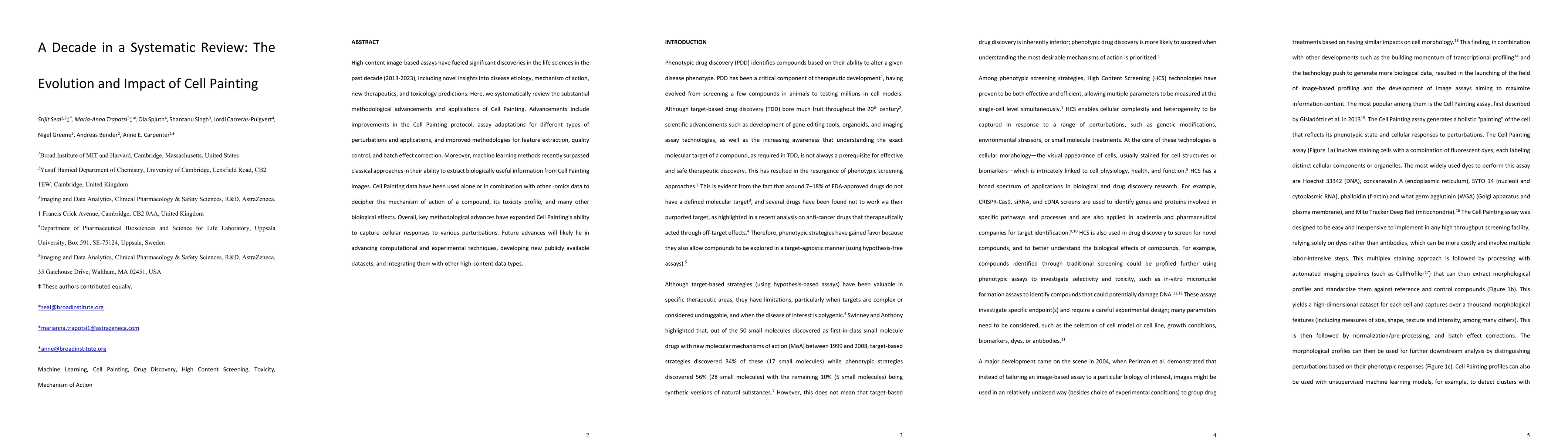
High-content image-based assays have fueled significant discoveries in the life sciences in the past decade (2013-2023), including novel insights into disease etiology, mechanism of action, new therapeutics, and toxicology predictions. Here, we systematically review the substantial methodological advancements and applications of Cell Painting. Advancements include improvements in the Cell Painting protocol, assay adaptations for different types of perturbations and applications, and improved methodologies for feature extraction, quality control, and batch effect correction. Moreover, machine learning methods recently surpassed classical approaches in their ability to extract biologically useful information from Cell Painting images. Cell Painting data have been used alone or in combination with other -omics data to decipher the mechanism of action of a compound, its toxicity profile, and many other biological effects. Overall, key methodological advances have expanded the ability of Cell Painting to capture cellular responses to various perturbations. Future advances will likely lie in advancing computational and experimental techniques, developing new publicly available datasets, and integrating them with other high-content data types.
AI Key Findings
Get AI-generated insights about this paper's methodology, results, significance, and more — seven facets brought into focus.
Paper Details
Authors
PDF Preview
Key Terms
Citation Network
Current paper (gray), citations (green), references (blue)
Display is limited for performance on very large graphs.
Related Papers
No references found for this paper.
Discussion 0