A Deep Learning Framework for Automatic Diagnosis in Lung Cancer
Publication
Metrics
AI Quick Summary
This paper presents a deep learning framework for automatically identifying and segmenting lung cancer areas in tissue specimens. The framework was trained and evaluated on tissue micro-arrays from 890 patients, achieving a pixel-wise precision of 0.80 and recall of 0.86, and demonstrated high performance on additional publicly available data.
Paper Preview
Abstract
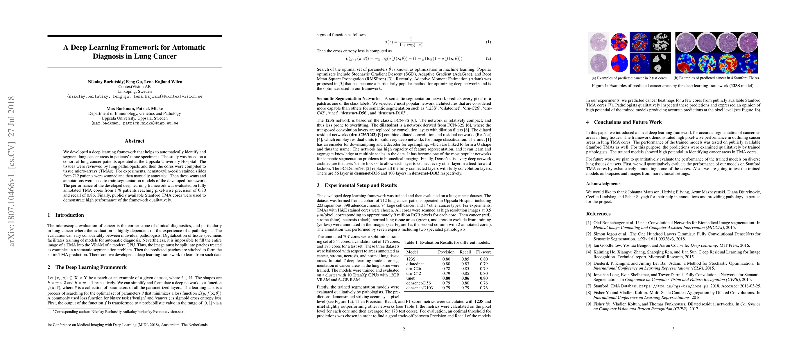
We developed a deep learning framework that helps to automatically identify and segment lung cancer areas in patients' tissue specimens. The study was based on a cohort of lung cancer patients operated at the Uppsala University Hospital. The tissues were reviewed by lung pathologists and then the cores were compiled to tissue micro-arrays (TMAs). For experiments, hematoxylin-eosin stained slides from 712 patients were scanned and then manually annotated. Then these scans and annotations were used to train segmentation models of the developed framework. The performance of the developed deep learning framework was evaluated on fully annotated TMA cores from 178 patients reaching pixel-wise precision of 0.80 and recall of 0.86. Finally, publicly available Stanford TMA cores were used to demonstrate high performance of the framework qualitatively.
AI Key Findings
Get AI-generated insights about this paper's methodology, results, significance, and more — seven facets brought into focus.
Impact
Paper Details
PDF Preview
Key Terms
Citation Network
Current paper (gray), citations (green), references (blue)
Display is limited for performance on very large graphs.
Discussion 0