ArxivLens

Quick Summary:

This paper introduces a deep convolutional neural network for classifying burn depths using ultrasound imaging, achieving 99% accuracy in identifying deep-partial thickness burns. The model learns from unburned and burned porcine skin samples, demonstrating high sensitivity and specificity, and shows potential for clinical use in burn depth assessment.

DOI: 10.1016/j.jmbbm.2021.104930
arXiv Id: 2203.15879
Date Published: 2022-03-31
Date Updated: 2022-03-31

Categories:

Key Terms (TF-IDF):

skin (0.310)
ultrasound (0.263)
classifier (0.229)
clinical (0.227)
classifier (0.226)
images (0.223)
encoder (0.223)
clinical (0.212)
decoder (0.204)
encoder (0.203)
images (0.198)
encoder decoder (0.187)
mode (0.181)
classify (0.173)
decoder (0.173)
classify (0.172)
mode (0.161)
depth (0.155)
accuracy (0.152)
trained (0.142)
License: http://arxiv.org/licenses/nonexclusive-distrib/1.0/

Summary:

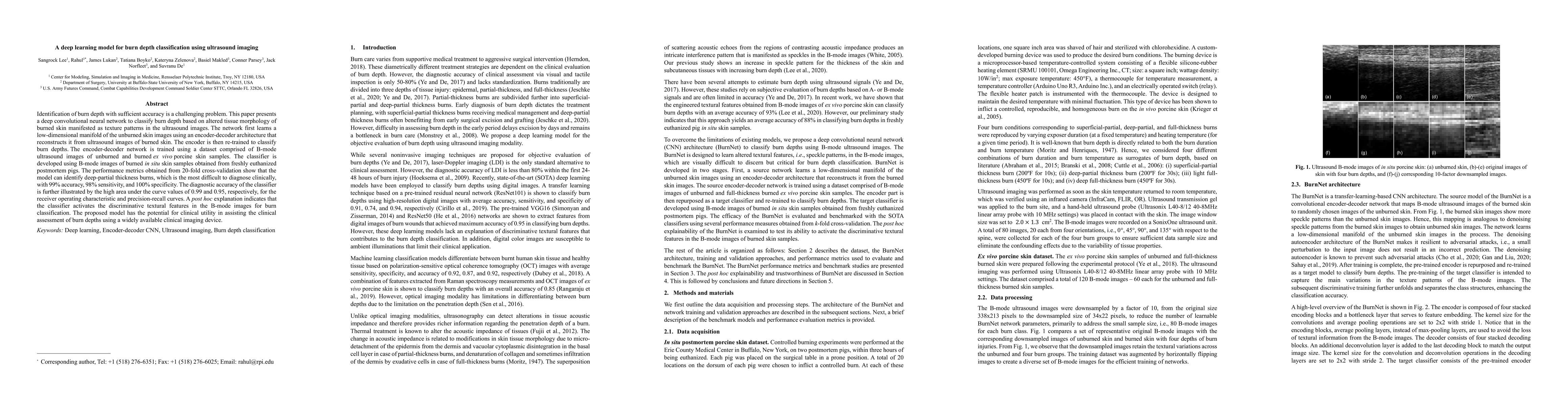
Identification of burn depth with sufficient accuracy is a challenging problem. This paper presents a deep convolutional neural network to classify burn depth based on altered tissue morphology of burned skin manifested as texture patterns in the ultrasound images. The network first learns a low-dimensional manifold of the unburned skin images using an encoder-decoder architecture that reconstructs it from ultrasound images of burned skin. The encoder is then re-trained to classify burn depths. The encoder-decoder network is trained using a dataset comprised of B-mode ultrasound images of unburned and burned ex vivo porcine skin samples. The classifier is developed using B-mode images of burned in situ skin samples obtained from freshly euthanized postmortem pigs. The performance metrics obtained from 20-fold cross-validation show that the model can identify deep-partial thickness burns, which is the most difficult to diagnose clinically, with 99% accuracy, 98% sensitivity, and 100% specificity. The diagnostic accuracy of the classifier is further illustrated by the high area under the curve values of 0.99 and 0.95, respectively, for the receiver operating characteristic and precision-recall curves. A post hoc explanation indicates that the classifier activates the discriminative textural features in the B-mode images for burn classification. The proposed model has the potential for clinical utility in assisting the clinical assessment of burn depths using a widely available clinical imaging device.

Citation & Reference Network Visualization

This graph shows how this paper is connected to other research. The current paper is shown in dark gray, papers that cite it are in green, and papers it references are in blue. You can:

Current Paper
Papers citing this work (Citations)
Papers cited by this work (References)

• Hover over any node to see the paper title

• Click a node to view that paper's details

• Drag nodes to rearrange the graph

• Use the zoom controls (bottom right) to zoom in/out

Citations and References

Title Authors Year Actions
Strategies for Optimizing Acute Burn Wound Therapy: A Comprehensive Review
A. Cretu, Andreea Grosu-Bularda, E. Bordeanu-Diaconescu, F. Hodea, Vlad-Alin Ratoiu, Catalina-Stefania Dumitru, M. Andrei, T. Neagu, I. Lascar, C. Hariga
2025
View Paper
WFUMB Commentary Paper on Artificial intelligence in Medical Ultrasound Imaging.
X. Cui, A. Goudie, Michael Blaivas, Young Jun Chai, M. Chammas, Yi Dong, Jonathon Stewart, Tian-an Jiang, Ping Liang, Chandra M Sehgal, Xing-Long Wu, Peter Ching-Chang Hsieh, Săftoiu Adrian, C. F. Dietrich
2024
View Paper
Detection of Human Skin Burn using ABNet Model
Priya Mittal, Bhisham Sharma, D. P. Yadav
2024
View Paper
Prediction of single cell mechanical properties in microchannels based on deep learning
Jiajie Gong, Xinyue Liu, Yancong Zhang, Fengping Zhu, Guohui Hu
2024
View Paper
Multi-Feature Extraction and Selection Method to Diagnose Burn Depth from Burn Images
Xizhe Zhang, Qi Zhang, Peixian Li, Jie You, Jingzhang Sun, Jianhang Zhou
2024
View Paper
Review of machine learning for optical imaging of burn wound severity assessment
Robert H Wilson, Rebecca Rowland, Gordon T. Kennedy, Chris Campbell, Victor C. Joe, Theresa L Chin, D. Burmeister, Robert J Christy, A. Durkin
2024
View Paper
Dense Mesh RCNN: assessment of human skin burn and burn depth severity
C. Pabitha, B. Vanathi
2023
View Paper
Ultrasonography for Skin and Soft Tissue Infections, Noninfectious Cysts, Foreign Bodies, and Burns in the Critical Care Setting.
Kelli A Craven, Kyle Luckey-Smith, Susanna Rudy
2023
View Paper
Spatial attention-based residual network for human burn identification and classification
D. P. Yadav, Turki Aljrees, Deepak Kumar, Ankit Kumar, K. Singh, Teekam Singh
2023
View Paper
Inhalation Injury Grading Using Transfer Learning Based on Bronchoscopy Images and Mechanical Ventilation Period
Yifan Li, Alan W. Pang, Jad F. Zeitouni, Ferris Zeitouni, Kirby Mateja, John A. Griswold, J. Chong
2022
View Paper
Enhancing Burn Severity Assessment With Deep Learning: A Comparative Analysis and Computational Efficiency Evaluation
Saeka Rahman, Md Motiur Rahman, M. Faezipour, Mo Rastgaar, Elika Ridelman, Justin D. Klein, Beth A. Angst, C. Shanti
2024
View Paper

Similar Papers

Human-centered XAI for Burn Depth Characterization

Juan Wachs, Yexiang Xue, Maria Romeo Tricas, Maxwell J. Jacobson, Daniela Chanci Arrubla, Gayle Gordillo, Chandan Sen

Published: 2023-01-03

Deep Learning-based Synthetic High-Resolution In-Depth Imaging Using an Attachable Dual-element Endoscopic Ultrasound Probe

Jihun Kim, Jaegeun Park, Hah Min Lew, Jae Seong Kim, Moon Hwan Lee, Sangyeon Youn, Hee Man Kim, Jae Youn Hwang

Published: 2023-09-14

Comments (0)

This website uses cookies to ensure you get the best experience. Learn more