A Deep Retinal Image Quality Assessment Network with Salient Structure Priors
Publication
Metrics
AI Quick Summary
This paper proposes SalStructuIQA, a deep retinal image quality assessment network that incorporates salient structure priors to focus on key anatomical features. The method includes two CNN architectures: Dual-branch SalStructIQA, which separately assesses large-size and tiny-size salient structures, and Single-branch SalStructIQA, which combines both structures. Experimental results show that Dual-branch SalStructIQA outperforms existing methods, while Single-branch offers a lightweight alternative with competitive performance.
Paper Preview
Abstract
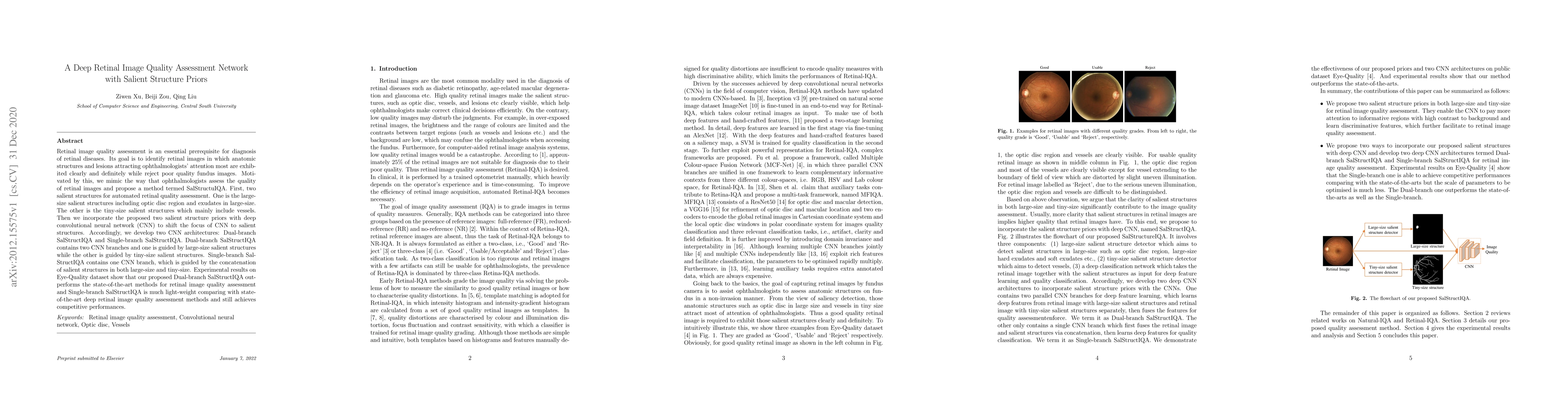
Retinal image quality assessment is an essential prerequisite for diagnosis of retinal diseases. Its goal is to identify retinal images in which anatomic structures and lesions attracting ophthalmologists' attention most are exhibited clearly and definitely while reject poor quality fundus images. Motivated by this, we mimic the way that ophthalmologists assess the quality of retinal images and propose a method termed SalStructuIQA. First, two salient structures for automated retinal quality assessment. One is the large-size salient structures including optic disc region and exudates in large-size. The other is the tiny-size salient structures which mainly include vessels. Then we incorporate the proposed two salient structure priors with deep convolutional neural network (CNN) to shift the focus of CNN to salient structures. Accordingly, we develop two CNN architectures: Dual-branch SalStructIQA and Single-branch SalStructIQA. Dual-branch SalStructIQA contains two CNN branches and one is guided by large-size salient structures while the other is guided by tiny-size salient structures. Single-branch SalStructIQA contains one CNN branch, which is guided by the concatenation of salient structures in both large-size and tiny-size. Experimental results on Eye-Quality dataset show that our proposed Dual-branch SalStructIQA outperforms the state-of-the-art methods for retinal image quality assessment and Single-branch SalStructIQA is much light-weight comparing with state-of-the-art deep retinal image quality assessment methods and still achieves competitive performances.
AI Key Findings
Get AI-generated insights about this paper's methodology, results, significance, and more — seven facets brought into focus.
Impact
Paper Details
Authors
PDF Preview
Key Terms
Citation Network
Current paper (gray), citations (green), references (blue)
Display is limited for performance on very large graphs.
Discussion 0