Summary

Capturing biological specimens at large scales with sub-micron resolution is crucial for biomedical research, but conventional cameras often can't handle the pixel requirements. While most microscopes use motorized stages to move samples and capture images tile by tile, we propose a method that eliminates the need for sample movement. Our approach integrates a scanning mechanism within the microscope's detection unit, enabling sequential capture of sub-areas of the field of view without physically moving the sample. This "remote scanning" method works with all camera-based microscopes and we demonstrate its effectiveness in both bright-field and epifluorescence microscopy. This technique is ideal for imaging motion-sensitive samples and large biological specimens, as evidenced in this study with millimeter-scale engineered tissues.

AI Key Findings

Generated Jun 10, 2025

Methodology

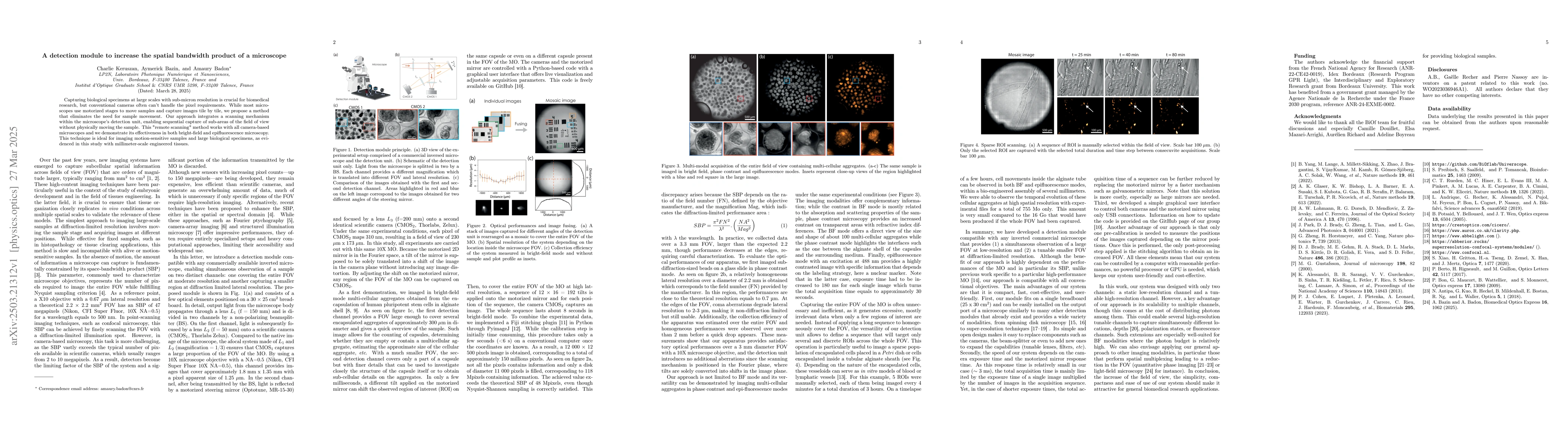
The research proposes a detection module integrated within a microscope's detection unit, enabling sequential capture of sub-areas of the field of view without physically moving the sample, termed 'remote scanning'. This method works with all camera-based microscopes and is demonstrated in bright-field and epifluorescence microscopy.

Key Results

  • The detection module allows for simultaneous observation of a large field of view (FOV) at low resolution and a tunable smaller FOV at diffraction-limited resolution.
  • The system achieves a higher spatial bandwidth product (SBP) than theoretical limits, surpassing 48Mpixels with Nyquist-Shannon sampling correctly satisfied.
  • The module is compatible with all conventional objectives and provides satisfactory optical performance over a 3mm diameter FOV with a 10X microscope objective.
  • The approach enables imaging of multi-cellular aggregates in bright-field, phase-contrast, and epifluorescence modes, offering complementary information.
  • The system's versatility allows targeting specific regions of interest (ROIs) across the FOV, reducing unnecessary data capture.

Significance

This research is significant as it presents a compact, fast, cost-effective, and user-friendly detection module for microscopy, enhancing the imaging of large biological specimens without sample movement. The method is particularly beneficial for motion-sensitive samples and large-scale biomedical research.

Technical Contribution

The main technical contribution is the development of a detection module that fits on a single breadboard, is easily installable on commercial microscopes, and offers a wide variety of modalities. Its simple and open design allows for easy replacement or addition of components.

Novelty

This work stands out for its compatibility with all conventional objectives, unlike previous work specific to high-performance microscopes. The system's compactness, speed, cost-effectiveness, and user-friendliness are key novel aspects.

Limitations

  • The method's benefits are highly dependent on the performance of the microscope, especially its SBP.
  • The system's acquisition time is mainly limited by the exposure time of individual images and the number of images in the sequence.
  • For faster acquisition, a more costly solution like galvanometric mirrors could be considered.

Future Work

  • Explore the scalability of the approach to multiple high-resolution tunable channels for simultaneous imaging of different locations, depths, polarization states, or fluorescence channels.
  • Apply the general approach to other imaging modalities that perform spatial multiplexing, leading to a reduction in the FOV, such as quantitative phase imaging or light-field microscopy.

Paper Details

PDF Preview

Download PDF

No citations found for this paper.

Comments (0)

This website uses cookies to ensure you get the best experience. Learn more