A dynamic graph-cuts method with integrated multiple feature maps for segmenting kidneys in ultrasound images
Publication
Metrics
AI Quick Summary
This paper proposes a dynamic graph-cuts method integrating multiple feature maps for kidney segmentation in ultrasound images, achieving superior results with an average Dice index of 0.9581, compared to other state-of-the-art methods. The method dynamically updates a localized graph to handle large appearance variations and weak boundaries.
Paper Preview
Abstract
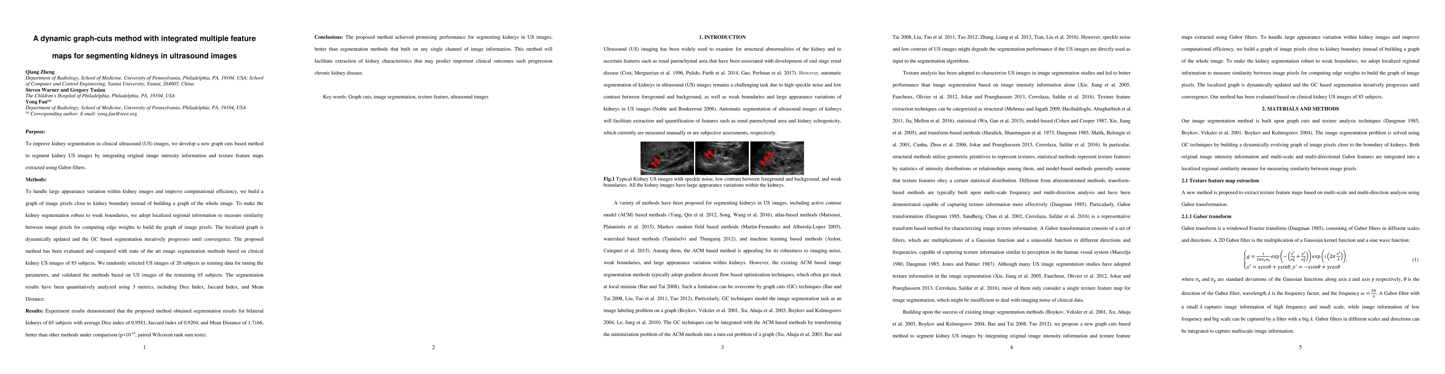
Purpose: To improve kidney segmentation in clinical ultrasound (US) images, we develop a new graph cuts based method to segment kidney US images by integrating original image intensity information and texture feature maps extracted using Gabor filters. Methods: To handle large appearance variation within kidney images and improve computational efficiency, we build a graph of image pixels close to kidney boundary instead of building a graph of the whole image. To make the kidney segmentation robust to weak boundaries, we adopt localized regional information to measure similarity between image pixels for computing edge weights to build the graph of image pixels. The localized graph is dynamically updated and the GC based segmentation iteratively progresses until convergence. The proposed method has been evaluated and compared with state of the art image segmentation methods based on clinical kidney US images of 85 subjects. We randomly selected US images of 20 subjects as training data for tuning the parameters, and validated the methods based on US images of the remaining 65 subjects. The segmentation results have been quantitatively analyzed using 3 metrics, including Dice Index, Jaccard Index, and Mean Distance. Results: Experiment results demonstrated that the proposed method obtained segmentation results for bilateral kidneys of 65 subjects with average Dice index of 0.9581, Jaccard index of 0.9204, and Mean Distance of 1.7166, better than other methods under comparison (p<10-19, paired Wilcoxon rank sum tests). Conclusions: The proposed method achieved promising performance for segmenting kidneys in US images, better than segmentation methods that built on any single channel of image information. This method will facilitate extraction of kidney characteristics that may predict important clinical outcomes such progression chronic kidney disease.
AI Key Findings
Get AI-generated insights about this paper's methodology, results, significance, and more — seven facets brought into focus.
Impact
Paper Details
PDF Preview
Key Terms
Citation Network
Current paper (gray), citations (green), references (blue)
Display is limited for performance on very large graphs.
Discussion 0