A Flexible 2.5D Medical Image Segmentation Approach with In-Slice and Cross-Slice Attention
Publication
Metrics
AI Quick Summary
This paper proposes CSA-Net, a flexible 2.5D medical image segmentation model that combines in-slice self-attention and cross-slice attention mechanisms to capture both local and global spatial relationships in 2.5D images. The model outperforms existing 2D and 2.5D segmentation methods in multi-class and binary segmentation tasks for brain and prostate MRIs.
Paper Preview
Abstract
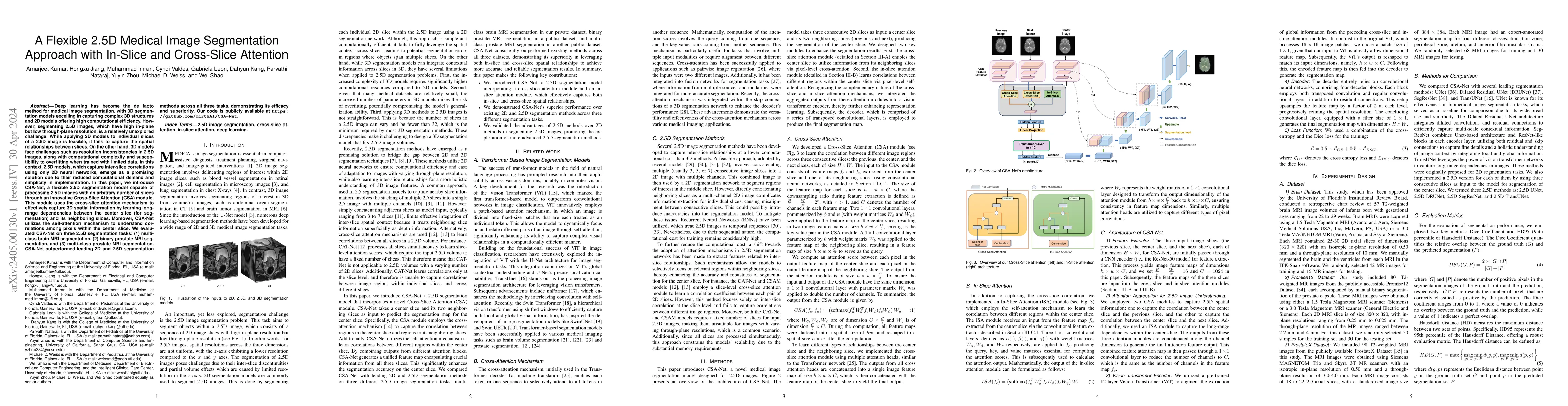
Deep learning has become the de facto method for medical image segmentation, with 3D segmentation models excelling in capturing complex 3D structures and 2D models offering high computational efficiency. However, segmenting 2.5D images, which have high in-plane but low through-plane resolution, is a relatively unexplored challenge. While applying 2D models to individual slices of a 2.5D image is feasible, it fails to capture the spatial relationships between slices. On the other hand, 3D models face challenges such as resolution inconsistencies in 2.5D images, along with computational complexity and susceptibility to overfitting when trained with limited data. In this context, 2.5D models, which capture inter-slice correlations using only 2D neural networks, emerge as a promising solution due to their reduced computational demand and simplicity in implementation. In this paper, we introduce CSA-Net, a flexible 2.5D segmentation model capable of processing 2.5D images with an arbitrary number of slices through an innovative Cross-Slice Attention (CSA) module. This module uses the cross-slice attention mechanism to effectively capture 3D spatial information by learning long-range dependencies between the center slice (for segmentation) and its neighboring slices. Moreover, CSA-Net utilizes the self-attention mechanism to understand correlations among pixels within the center slice. We evaluated CSA-Net on three 2.5D segmentation tasks: (1) multi-class brain MRI segmentation, (2) binary prostate MRI segmentation, and (3) multi-class prostate MRI segmentation. CSA-Net outperformed leading 2D and 2.5D segmentation methods across all three tasks, demonstrating its efficacy and superiority. Our code is publicly available at https://github.com/mirthAI/CSA-Net.
AI Key Findings
Get AI-generated insights about this paper's methodology, results, significance, and more — seven facets brought into focus.
Impact
Paper Details
Authors
PDF Preview
Key Terms
Citation Network
Current paper (gray), citations (green), references (blue)
Display is limited for performance on very large graphs.
Discussion 0