A flexible semi-automatic approach for segmenting glioblastoma multiforme in MRI images achieved an average Dice Similarity Coefficient of 77.72% with user-defined seed points, outperforming a one-click method.
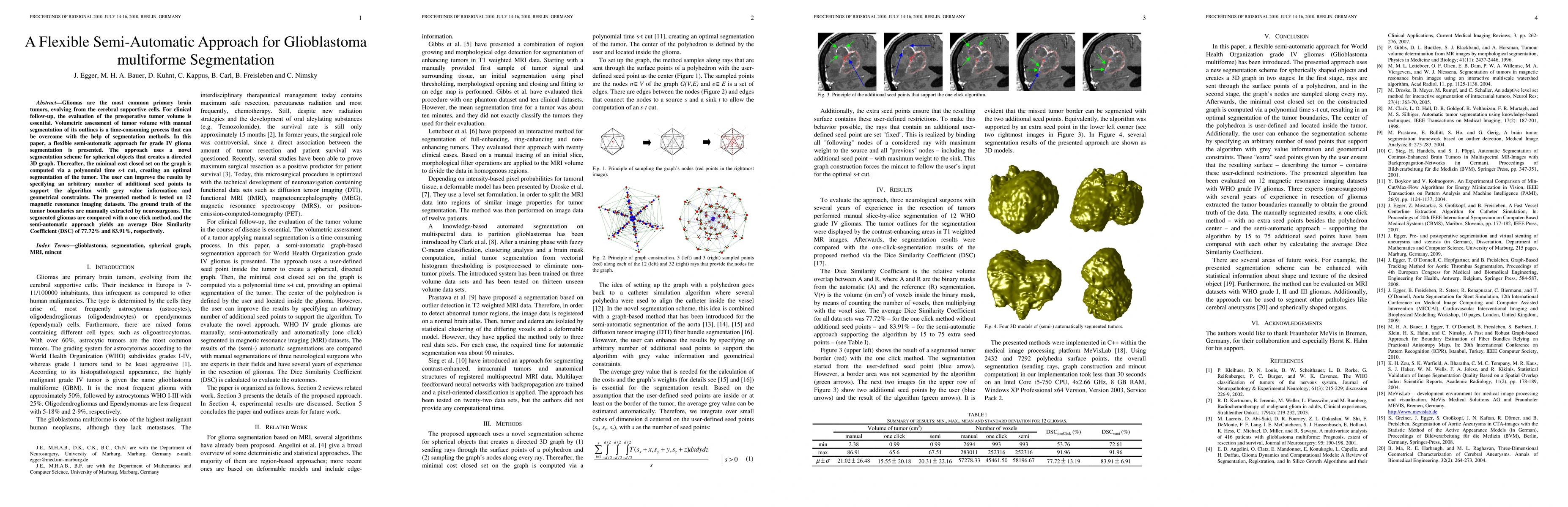
Gliomas are the most common primary brain tumors, evolving from the cerebral supportive cells. For clinical follow-up, the evaluation of the preoperative tumor volume is essential. Volumetric assessment of tumor volume with manual segmentation of its outlines is a time-consuming process that can be overcome with the help of segmentation methods. In this paper, a flexible semi-automatic approach for grade IV glioma segmentation is presented. The approach uses a novel segmentation scheme for spherical objects that creates a directed 3D graph. Thereafter, the minimal cost closed set on the graph is computed via a polynomial time s-t cut, creating an optimal segmentation of the tumor. The user can improve the results by specifying an arbitrary number of additional seed points to support the algorithm with grey value information and geometrical constraints. The presented method is tested on 12 magnetic resonance imaging datasets. The ground truth of the tumor boundaries are manually extracted by neurosurgeons. The segmented gliomas are compared with a one click method, and the semi-automatic approach yields an average Dice Similarity Coefficient (DSC) of 77.72% and 83.91%, respectively.
Get AI-generated insights about this paper's methodology, results, and significance.
Current paper (gray), citations (green), references (blue)
Title | Authors | Year | Actions |
---|---|---|---|
Lower jawbone data generation for deep learning tools under MeVisLab
|
Birgit Pfarrkirchner, Christina Gsaxner, L. Lindner, N. Jakse, J. Wallner, D. Schmalstieg, J. Egger
|
2018
|
View Paper |
Multi-threaded integration of HTC-Vive and MeVisLab
|
Simon Gunacker, Markus Gall, D. Schmalstieg, J. Egger
|
2018
|
View Paper |
In-depth assessment of an interactive graph-based approach for the segmentation for pancreatic metastasis in ultrasound acquisitions of the liver with two specialists in Internal Medicine
|
J. Egger, Xiaojun Chen, L. Bettac, M. Hänle, T. Gräter, W. Zoller, D. Schmalstieg, A. Hann
|
2017
|
View Paper |
Integration of the HTC Vive into the medical platform MeVisLab
|
J. Egger, Markus Gall, J. Wallner, Pedro de Almeida Germano Boechat, A. Hann, Xing Li, Xiaojun Chen, D. Schmalstieg
|
2017
|
View Paper |
Algorithmic evaluation of lower jawbone segmentations
|
J. Egger, Kerstin Hochegger, Markus Gall, Xiaojun Chen, K. Reinbacher, K. Schwenzer-Zimmerer, D. Schmalstieg, J. Wallner
|
2017
|
View Paper |
Interactive planning of miniplates
|
Markus Gall, K. Reinbacher, J. Wallner, Jan Stanzel, Xiaojun Chen, K. Schwenzer-Zimmerer, D. Schmalstieg, J. Egger
|
2017
|
View Paper |
Computer-aided planning and reconstruction of cranial 3D implants
|
Markus Gall, Xing Li, Xiaojun Chen, D. Schmalstieg, J. Egger
|
2016
|
View Paper |
US-Cut: interactive algorithm for rapid detection and segmentation of liver tumors in ultrasound acquisitions
|
J. Egger, Philip Voglreiter, Mark Dokter, M. Hofmann, Xiaojun Chen, W. Zoller, D. Schmalstieg, A. Hann
|
2016
|
View Paper |
Cellular automata segmentation of the boundary between the compacta of vertebral bodies and surrounding structures
|
J. Egger, C. Nimsky
|
2016
|
View Paper |
Interactive and scale invariant segmentation of the rectum/sigmoid via user-defined templates
|
Tobias Lüddemann, J. Egger
|
2016
|
View Paper |
Preoperative volume determination for pituitary adenoma
|
Dženan Zukić, J. Egger, M. Bauer, D. Kuhnt, B. Carl, Bernd Freisleben, A. Kolb, C. Nimsky
|
2016
|
View Paper |
Segmentation of Glioblastoma Multiforme from MR Images – A comprehensive review
|
V. R. Simi, J. Joseph
|
2015
|
View Paper |
RFA-cut: Semi-automatic segmentation of radiofrequency ablation zones with and without needles via optimal s-t-cuts
|
J. Egger, H. Busse, P. Brandmaier, D. Seider, M. Gawlitza, S. Strocka, Philip Voglreiter, Mark Dokter, M. Hofmann, Bernhard Kainz, Xiaojun Chen, A. Hann, Pedro Boechat, Wei Yu, Bernd Freisleben, Tuomas Alhonnoro, M. Pollari, M. Moche, D. Schmalstieg
|
2015
|
View Paper |
Interactive Volumetry Of Liver Ablation Zones
|
J. Egger, H. Busse, P. Brandmaier, D. Seider, M. Gawlitza, S. Strocka, Philip Voglreiter, Mark Dokter, M. Hofmann, Bernhard Kainz, A. Hann, Xiaojun Chen, Tuomas Alhonnoro, M. Pollari, D. Schmalstieg, M. Moche
|
2015
|
View Paper |
An efficient inflation method for segmentation of medical 3D images
|
Dženan Zukić
|
2015
|
View Paper |
Differentiation between vasogenic-edema versus tumor-infiltrative area in patients with glioblastoma during bevacizumab therapy: a longitudinal MRI study.
|
M. Artzi, F. Bokstein, D. Blumenthal, O. Aizenstein, G. Liberman, B. Corn, D. Ben Bashat
|
2014
|
View Paper |
Refinement-Cut: User-Guided Segmentation Algorithm for Translational Science
|
J. Egger
|
2014
|
View Paper |
Interactive-cut: Real-time feedback segmentation for translational research
|
J. Egger, Tobias Lüddemann, Robert Schwarzenberg, Bernd Freisleben, C. Nimsky
|
2014
|
View Paper |
PCG-Cut: Graph Driven Segmentation of the Prostate Central Gland
|
J. Egger
|
2013
|
View Paper |
GBM Volumetry using the 3D Slicer Medical Image Computing Platform
|
J. Egger, T. Kapur, Andrey Fedorov, S. Pieper, James V. Miller, H. Veeraraghavan, Bernd Freisleben, A. Golby, C. Nimsky, R. Kikinis
|
2013
|
View Paper |
Manual Refinement System for Graph-Based Segmentation Results in the Medical Domain
|
J. Egger, R. Colen, Bernd Freisleben, C. Nimsky
|
2012
|
View Paper |
A Medical Software System for Volumetric Analysis of Cerebral Pathologies in Magnetic Resonance Imaging (MRI) Data
|
J. Egger, C. Kappus, Bernd Freisleben, C. Nimsky
|
2012
|
View Paper |
Square-Cut: A Segmentation Algorithm on the Basis of a Rectangle Shape
|
J. Egger, T. Kapur, T. Dukatz, M. Kolodziej, Dženan Zukić, Bernd Freisleben, C. Nimsky
|
2012
|
View Paper |
Pituitary Adenoma Segmentation
|
J. Egger, M. Bauer, D. Kuhnt, Bernd Freisleben, C. Nimsky
|
2011
|
View Paper |
A Comparison of Two Human Brain Tumor Segmentation Methods for MRI Data
|
J. Egger, Dženan Zukić, M. Bauer, D. Kuhnt, B. Carl, Bernd Freisleben, A. Kolb, C. Nimsky
|
2011
|
View Paper |
Glioblastoma Multiforme Segmentation in MRI Data with a Balloon Inflation Approach
|
Dženan Zukić, J. Egger, M. Bauer, D. Kuhnt, B. Carl, Bernd Freisleben, A. Kolb, C. Nimsky
|
2011
|
View Paper |
Nugget-Cut: A Segmentation Scheme for Spherically- and Elliptically-Shaped 3D Objects
|
J. Egger, M. Bauer, D. Kuhnt, B. Carl, C. Kappus, Bernd Freisleben, C. Nimsky
|
2010
|
View Paper |
Computer-aided planning and reconstruction of cranial 3D implants.
|
Markus Gall, Xing Li, Xiaojun Chen, D. Schmalstieg, J. Egger
|
2016
|
View Paper |
Semi-automatische Segmentierung von Schädigungszonen in post-interventionellen CT-Daten
|
J. Egger, H. Busse, M. Moche, P. Brandmaier, D. Seider, M. Gawlitza, S. Strocka, N. Garnov, J. Fuchs, P. Voigt, F. Dazinger, P. Voglreiter, M. Dokter, M. Hofmann, A. Hann, Bernd Freisleben, T. Kahn, D. Schmalstieg
|
2015
|
View Paper |
Registration and Segmentation for Image-Guided Therapy
|
T. Kapur, J. Egger, J. Jayender, M. Toews, W. Wells
|
2014
|
View Paper |
Semi-automatische Echtzeit-Konturierung
|
J. Egger
|
2014
|
View Paper |
Semi - automatische Echtzeit - Konturierung mit einem vorlagenbasierten und skalierungsinvarianten Ansatz
|
J. Egger
|
2014
|
View Paper |
Comments (0)