This paper proposes a fully convolutional neural network for retinal vessel segmentation, framing it as a multi-label inference task. The model significantly outperforms existing methods, achieving 95.33% accuracy and a 0.974 AUC score on the DRIVE dataset.
Automatic segmentation of retinal blood vessels from fundus images plays an important role in the computer aided diagnosis of retinal diseases. The task of blood vessel segmentation is challenging due to the extreme variations in morphology of the vessels against noisy background. In this paper, we formulate the segmentation task as a multi-label inference task and utilize the implicit advantages of the combination of convolutional neural networks and structured prediction. Our proposed convolutional neural network based model achieves strong performance and significantly outperforms the state-of-the-art for automatic retinal blood vessel segmentation on DRIVE dataset with 95.33% accuracy and 0.974 AUC score.
Generated Sep 02, 2025
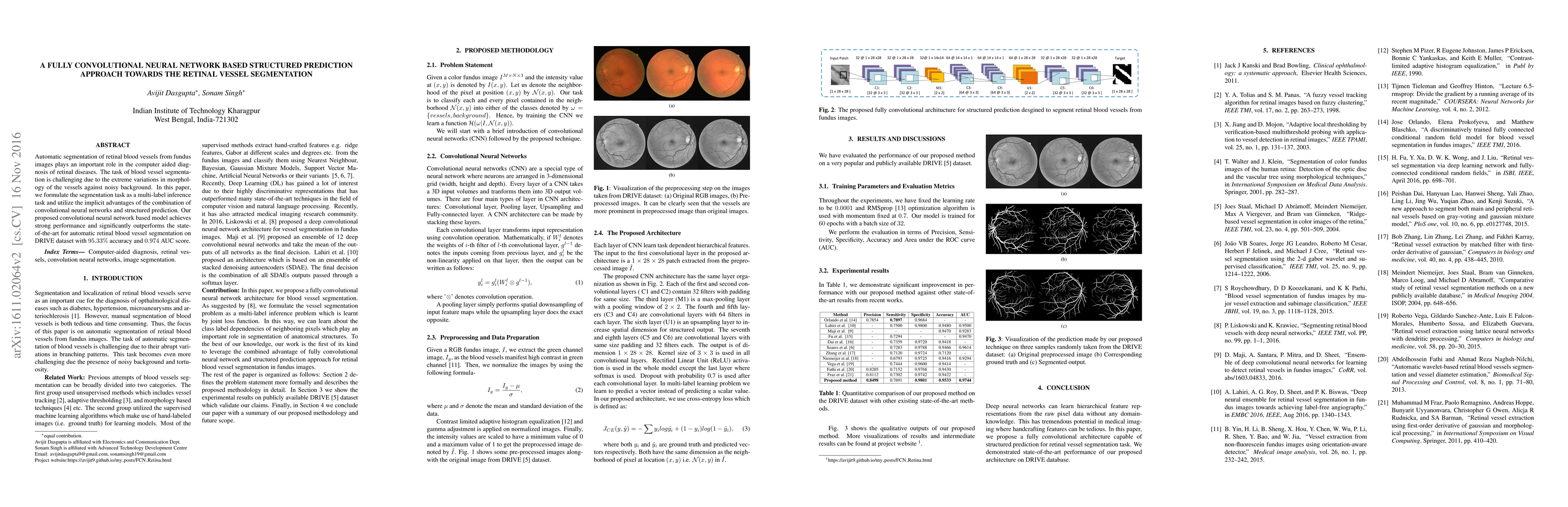
The paper proposes a fully convolutional neural network (FCN) architecture for retinal blood vessel segmentation, formulated as a multi-label inference problem. It utilizes structured prediction to learn class label dependencies of neighboring pixels, leveraging the combined advantages of FCNs and structured prediction for retinal blood vessel segmentation in fundus images.
This research is important as it presents a deep learning approach for automatic retinal blood vessel segmentation, which is crucial for computer-aided diagnosis of retinal diseases. The method reduces the need for manual, time-consuming segmentation, and offers high accuracy.
The paper introduces a novel FCN architecture for retinal vessel segmentation, incorporating structured prediction to capture dependencies between neighboring pixels, leading to improved segmentation performance.
The proposed method combines FCNs with structured prediction for retinal blood vessel segmentation, offering better performance compared to existing techniques, and is one of the first to leverage this combined approach in medical image segmentation.
Current paper (gray), citations (green), references (blue)
Display is limited for performance on very large graphs.
Tariq M Khan, Syed S. Naqvi, Antonio Robles-Kelly
Title | Authors | Year | Actions |
---|
Comments (0)