01
MethodologyHow they did it
The study employed a novel 3D bioprinting approach to create functional human liver tissue models.
This paper presents a scalable 3D bioprinting technique to create functional human liver tissue models in the form of discoid structures. The 3D printed liver tissue models exhibit superior albumin and urea synthesis, ADME gene expression, and metabolic activity compared to traditional spheroid models, showing potential for drug development applications.
The study employed a novel 3D bioprinting approach to create functional human liver tissue models. More in Methodology →
The co-culture discoid model exhibited improved hepatocyte functionality compared to traditional monolayer cultures. — Enhanced expression of detoxification genes and increased CYP2B6 activity were observed in the 3D bioprinted liver tissue. More in Key Results →
This research contributes to the development of more accurate and effective models for liver disease modeling and drug testing. More in Significance →
Limited sample size and short-term culture duration. — Inability to fully replicate human liver physiology in vitro. More in Limitations →
To reduce costs and delays related to developing new and effective drugs, there is a critical need for improved human liver tissue models. Here we describe an approach for 3D bioprinting functional human liver tissue models, in which we fabricate disc-shaped structures (discoids) 200 {\mu}m in thickness and 1-3 mm in diameter, embedded in a highly permeable support medium made from packed microgels. We demonstrate that the method is precise, accurate, and scalable; up to 100 tissues per hour can be manufactured with a variability and error in diameter of about 4%. Histologic and immunohistochemical evaluation of printed discs reveal self-organization, cell cohesion, and key liver marker expression. During the course of 3-4 weeks in culture, the tissues stably synthesize albumin and urea at high levels, outperforming spheroid tissue models. We find the tissues express more than 100 genes associated with molecular absorption, distribution, metabolism, and excretion (ADME) at levels within the range of human liver. The liver tissue models exhibit enzymatic formation of metabolites after exposure to multiple test compounds. Together, these results demonstrate the promise of 3D printed discoids for pharmacological and toxicological applications.
Seven facets of this paper, analysed and brought into focus by AI.
This research contributes to the development of more accurate and effective models for liver disease modeling and drug testing.
The study employed a novel 3D bioprinting approach to create functional human liver tissue models.
This research contributes to the development of more accurate and effective models for liver disease modeling and drug testing.
The development of a novel 3D bioprinting approach for creating functional human liver tissue models.
This study introduces a new paradigm for liver tissue engineering, offering potential applications in personalized medicine and drug discovery.
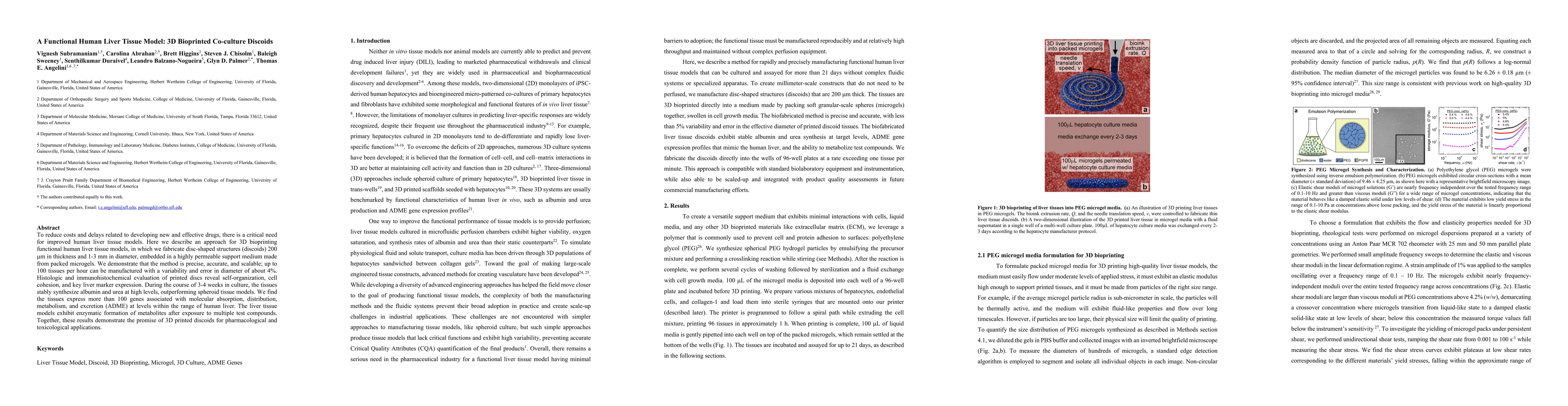
Current paper (gray), citations (green), references (blue)
Display is limited for performance on very large graphs.
Discussion 0