ArxivLens

Quick Summary:

This paper presents an optimized geometry for nanoscale magnetic resonance force microscopy, achieving high magnetic field gradients and minimal dissipation, which could enhance MRFM applications for low-gyro-magnetic nuclear species and broadened resonance samples. The experimental setup uses a cantilever aligned perpendicularly to both the magnetic field and RF source.

Journal Reference: Appl. Phys. Lett. 98, 163103 (2011)
DOI: 10.1063/1.3579521
arXiv Id: 1103.4241
Comments: 4 pages, 5 figures
Date Published: 2015-03-19
Date Updated: 2015-03-19

Key Terms (TF-IDF):

rf (0.314)
magnetic (0.288)
geometry (0.212)
tip (0.191)
mhz (0.179)
external magnetic (0.176)
dots (0.173)
resonances (0.171)
spins (0.168)
microscopy (0.164)
negligible (0.163)
facilitate (0.161)
gradients (0.160)
dissipation (0.160)
rotating (0.158)
species (0.158)
nm (0.157)
axis (0.155)
magnetic fields (0.152)
implement (0.147)
License: http://arxiv.org/licenses/nonexclusive-distrib/1.0/

Summary:

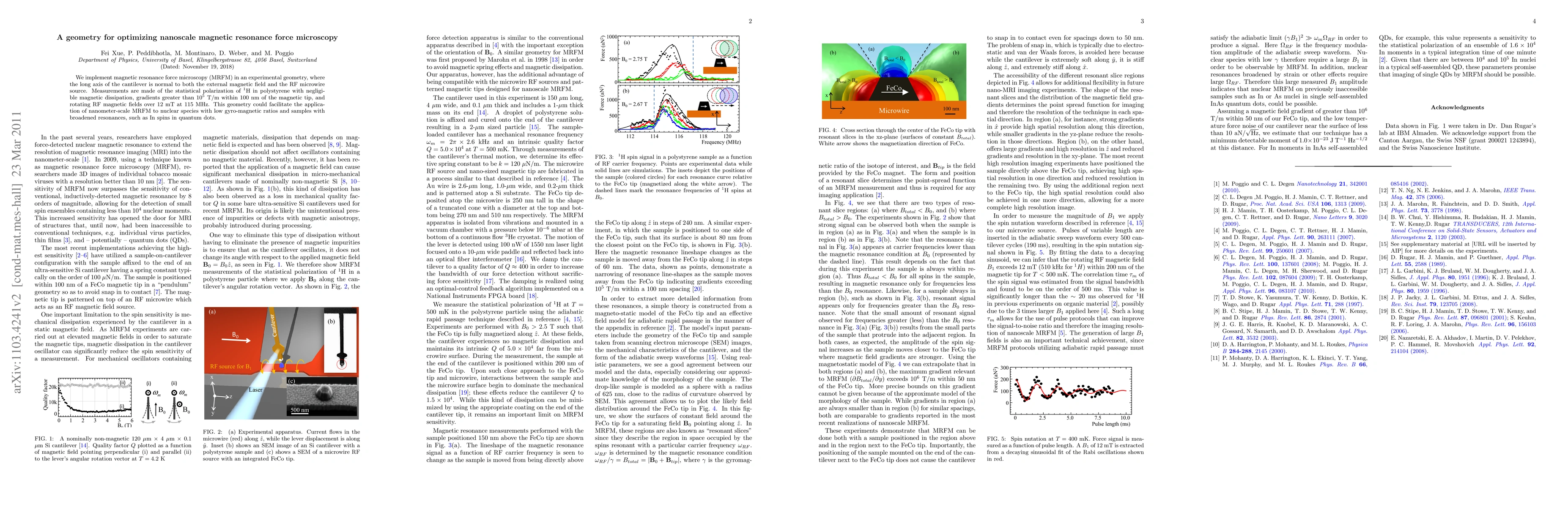
We implement magnetic resonance force microscopy (MRFM) in an experimental geometry, where the long axis of the cantilever is normal to both the external magnetic field and the RF microwire source. Measurements are made of the statistical polarization of $^1$H in polystyrene with negligible magnetic dissipation, gradients greater than $10^5$ T/m within 100 nm of the magnetic tip, and rotating RF magnetic fields over 12 mT at 115 MHz. This geometry could facilitate the application of nanometer-scale MRFM to nuclear species with low gyro-magnetic ratios and samples with broadened resonances, such as In spins in quantum dots.

Citation & Reference Network Visualization

This graph shows how this paper is connected to other research. The current paper is shown in dark gray, papers that cite it are in green, and papers it references are in blue. You can:

Current Paper
Papers citing this work (Citations)
Papers cited by this work (References)

• Hover over any node to see the paper title

• Click a node to view that paper's details

• Drag nodes to rearrange the graph

• Use the zoom controls (bottom right) to zoom in/out

Citations and References

Title Authors Year Actions
Visualisation and evaluation of electrical contact spots and asperities of a given contact system using x-ray computed tomography
C. Roussos
2018
View Paper
Force‐Detected Nuclear Magnetic Resonance
M. Poggio, B. Herzog
2017
View Paper
A 3D contact analysis approach for the visualization of the electrical contact asperities
C. Roussos, J. Swingler
2017
View Paper
High-Gradient Nanomagnet-On-Cantilever Fabrication For Scanned Probe Detection Of Magnetic Resonance
Jonilyn G. Longenecker
2013
View Paper
Cantilever magnetometry of individual Ni nanotubes.
D. Weber, D. Rüffer, A. Buchter, F. Xue, E. Russo-Averchi, R. Huber, P. Berberich, J. Arbiol, A. Fontcuberta i Morral, D. Grundler, M. Poggio
2012
View Paper
1. Mechanical Detection Of Electron Spin Resonance From Nitroxide Spin Probes, 2. Ultrasensitive Cantilever Torque Magnetometry Of Magnetization Switching In Individual Nickel Nanorods
E. Moore
2012
View Paper
Measurement of statistical nuclear spin polarization in a nanoscale GaAs sample
F. Xue, D. Weber, P. Peddibhotla, M. Poggio
2011
View Paper
References
2019
View Paper
Hybrid torque and SQUID magnetometry of individual magnetic nanotubes
A. Buchter
2015
View Paper
Coupling of nanomechanical resonators to controllable quantum systems
M. Montinaro, H. Riel, J. Schibler, V. D. Palumbo
2014
View Paper
Dynamic cantilever magnetometry of individual ferromagnetic nanotubes
D. Weber
2014
View Paper

Comments (0)

This website uses cookies to ensure you get the best experience. Learn more