Authors
Publication
Metrics
Quick Actions
Quick Answers
What methodology did the authors use?
Flow Induced Dispersion Analysis (FIDA) using intrinsic phenylalanine fluorescence in peak and front modes to monitor monomer to oligomer conversion under near‑physiological flow conditions. More in Methodology →
What are the key results?
Quantitative detection of early‑stage oligomers (2–10 nm) and their size increase during aggregation of PSMα3 and hIAPP. — Observation of a biphasic depletion of soluble species with an initial fast consumption followed by a slower phase, confirmed by gel electrophoresis and TEM. More in Key Results →
Why is this work significant?
Provides a label‑free, real‑time analytical framework that captures early toxic oligomers, overcoming limitations of ThT and other extrinsic assays and enabling better therapeutic target identification. More in Significance →
What are the main limitations?
Insoluble aggregates appear as spikes and cannot be quantified by Taylor dispersion, limiting detection of mature fibrils. — Peak‑mode requires relatively high peptide concentrations, which may alter aggregation pathways. More in Limitations →
Abstract
The aggregation of amyloid-forming peptides is a dynamic, complex process that underlies their diverse biological activities, from physiological functions to disease-associated dysfunctions. While the structure of fibrillar end-products is well-characterized for most amyloids, the heterogeneous and often transient oligomers, likely key in cytotoxicity, remain poorly investigated, especially for peptides with low-yield aromatic residues. Here, by exploiting and developing flow induced dispersion analysis in both peak and front modes, we demonstrate that intrinsic phenylalanine fluorescence can be harnessed to quantify the conversion of diffusing monomers into non-diffusing oligomers and fibrils. We further characterize low-molecular-weight oligomers, and their size evolution from 2 to 10 nm over time. Importantly, we validate the robustness of our approach using two tryptophan-free and fast-fibrillating amyloid peptides, PSM$α$3 and hIAPP, known for their key roles in S. aureus virulence and type 2 diabetes respectively. Our results overcome the limitations of traditional biochemical and biophysical amyloid assays by extending analysis from large oligomers and fibrils to small heterogeneous oligomers, under near-physiological conditions. This study thus offers a new analytical framework, thereby filling a critical gap in amyloid research, to probe the early stages of aggregation, key in the design of alternative therapeutics for amyloid-diseases.
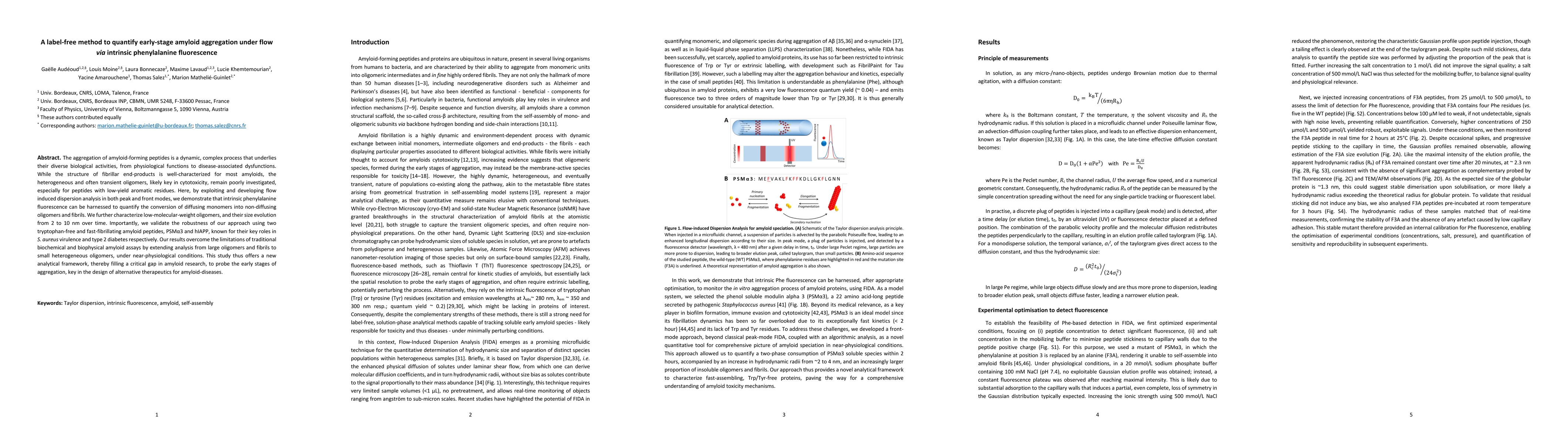
AI Key Findings
Generated Feb 24, 2026
Methodology — What approach did the authors take?
Flow Induced Dispersion Analysis (FIDA) using intrinsic phenylalanine fluorescence in peak and front modes to monitor monomer to oligomer conversion under near‑physiological flow conditions.
Key Results — What are the main findings?
- Quantitative detection of early‑stage oligomers (2–10 nm) and their size increase during aggregation of PSMα3 and hIAPP.
- Observation of a biphasic depletion of soluble species with an initial fast consumption followed by a slower phase, confirmed by gel electrophoresis and TEM.
- Demonstration that front‑mode FIDA allows analysis at low concentrations (50 µM) and yields a linear calibration curve for soluble species concentration.
Significance — Why does this research matter?
Provides a label‑free, real‑time analytical framework that captures early toxic oligomers, overcoming limitations of ThT and other extrinsic assays and enabling better therapeutic target identification.
Technical Contribution — What is the technical contribution?
Implementation of a constrained regularized linear inversion (CRLI) algorithm for size distribution extraction from peak and front mode Taylorgrams, enabling quantitative hydrodynamic radius determination of heterogeneous oligomers.
Novelty — What is new about this work?
First quantitative use of intrinsic phenylalanine fluorescence for amyloid aggregation analysis and the novel application of front‑mode FIDA to low‑concentration, fast‑fibrillating peptides.
Limitations — What are the limitations of this study?
- Insoluble aggregates appear as spikes and cannot be quantified by Taylor dispersion, limiting detection of mature fibrils.
- Peak‑mode requires relatively high peptide concentrations, which may alter aggregation pathways.
Future Work — What did the authors propose for future work?
- Develop a theoretical model to analyze spike signals and quantify large insoluble species.
- Integrate FIDA with complementary techniques (ThT, TEM, AFM) for a comprehensive speciation map.
Paper Details
How to Cite This Paper
@article{audéoud2026a,
title = {A label-free method to quantify early-stage amyloid aggregation under flow via intrinsic phenylalanine fluorescence},
author = {Audéoud, Gaëlle and Bonnecaze, Laura and Moine, Louis and others},
year = {2026},
eprint = {2602.19817},
archivePrefix = {arXiv},
primaryClass = {cond-mat.soft},
}
Audéoud, G., Bonnecaze, L., Moine, L., Khemtemourian, L., Mathelié-Guinlet, M., Lavaud, M., Salez, T., & Amarouchene, Y. (2026). A label-free method to quantify early-stage amyloid aggregation under flow via intrinsic phenylalanine fluorescence. arXiv. https://arxiv.org/abs/2602.19817
Audéoud, Gaëlle, et al. "A label-free method to quantify early-stage amyloid aggregation under flow via intrinsic phenylalanine fluorescence." arXiv, 2026, arxiv.org/abs/2602.19817.
PDF Preview
Similar Papers
Found 4 papersIntrinsic visible emission of amyloid-β oligomers: a potential tool for early alzheimer's diagnosis.
Novo, Mercedes, Illodo, Sara, Seijas, Jesús et al.
FLARE: a label-free fluorescence-assisted method for RNA engineering of three-way junctions.
Sharma, Ashwani, Krishna, A Murali, Fathima, Nida et al.
Label Differential Privacy via Aggregation
Anand Brahmbhatt, Rishi Saket, Shreyas Havaldar et al.
A Model-Free Method to Quantify Memory Utilization in Neural Point Processes
Gorana Mijatovic, Sebastiano Stramaglia, Luca Faes
Comments (0)