Publication
Metrics
Quick Actions
AI Quick Summary
This paper proposes a lightweight, extensible model for cell segmentation and classification in whole slide images, achieving improved data quality and performance through refined annotations and knowledge distillation from a foundation model. Integration into the QuPath platform shows significant gains in segmentation accuracy and efficiency, potentially reducing pathologist workload and enhancing diagnostic outcomes.
Quick Answers
What methodology did the authors use?
The research proposes a lightweight and extensible cell segmentation and classification model for whole slide images, addressing challenges in digital pathology by refining annotations, leveraging foundation models, distilling knowledge to reduce model size, and integrating the model into QuPath. More in Methodology →
What are the key results?
Improved segmentation and classification performance using the H-Optimus-based model compared to a CNN-based model, with average R^2 improving from 0.575 to 0.871 and average PQ score from 0.450 to 0.492. — The distilled model maintains comparable performance while reducing parameter count by a factor of 48. More in Key Results →
Why is this work significant?
This research is significant as it tackles critical challenges in digital pathology, such as dataset granularity, inconsistent annotations, high computational demands, and difficulties integrating new technologies into workflows, which could significantly impact clinical diagnostics. More in Significance →
What are the main limitations?
The method shows promise but requires extensive validation prior to clinical deployment. — Lower performance in detecting macrophages was observed, highlighting the broader challenge of accurately identifying complex cell types. More in Limitations →
Abstract
Developing clinically useful cell-level analysis tools in digital pathology remains challenging due to limitations in dataset granularity, inconsistent annotations, high computational demands, and difficulties integrating new technologies into workflows. To address these issues, we propose a solution that enhances data quality, model performance, and usability by creating a lightweight, extensible cell segmentation and classification model. First, we 
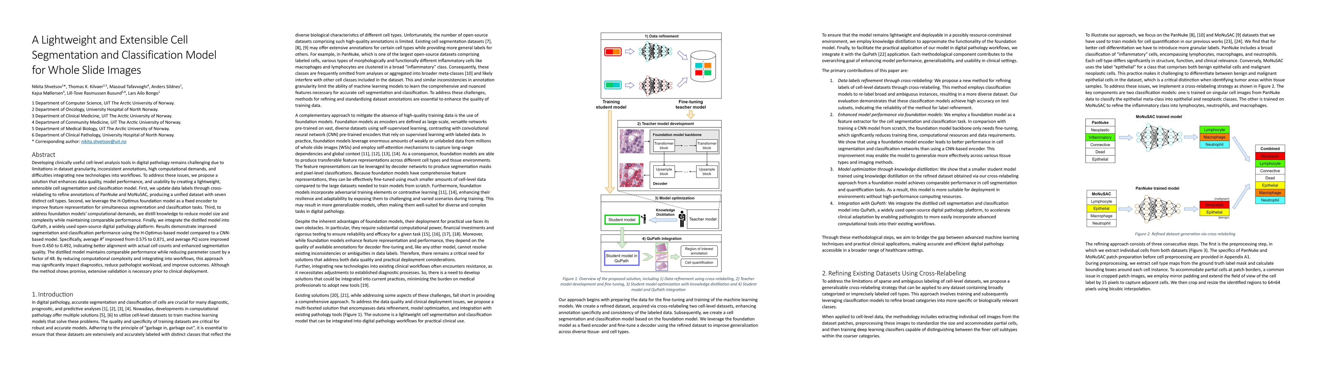
update data labels through cross-relabeling to refine annotations of PanNuke and MoNuSAC, producing a unified dataset with seven distinct cell types. Second, we leverage the H-Optimus foundation model as a fixed encoder to improve feature representation for simultaneous segmentation and classification tasks. Third, to address foundation models' computational demands, we distill knowledge to reduce model size and complexity while maintaining comparable performance. Finally, we integrate the distilled model into QuPath, a widely used open-source digital pathology platform. Results demonstrate improved segmentation and classification performance using the H-Optimus-based model compared to a CNN-based model. Specifically, average $R^2$ improved from 0.575 to 0.871, and average $PQ$ score improved from 0.450 to 0.492, indicating better alignment with actual cell counts and enhanced segmentation quality. The distilled model maintains comparable performance while reducing parameter count by a factor of 48. By reducing computational complexity and integrating into workflows, this approach may significantly impact diagnostics, reduce pathologist workload, and improve outcomes. Although the method shows promise, extensive validation is necessary prior to clinical deployment.
AI Key Findings
Generated Jun 11, 2025
Methodology — What approach did the authors take?
The research proposes a lightweight and extensible cell segmentation and classification model for whole slide images, addressing challenges in digital pathology by refining annotations, leveraging foundation models, distilling knowledge to reduce model size, and integrating the model into QuPath.
Key Results — What are the main findings?
- Improved segmentation and classification performance using the H-Optimus-based model compared to a CNN-based model, with average R^2 improving from 0.575 to 0.871 and average PQ score from 0.450 to 0.492.
- The distilled model maintains comparable performance while reducing parameter count by a factor of 48.
- Integration of the distilled model into QuPath, a widely used open-source digital pathology platform, demonstrates potential for impacting diagnostics, reducing pathologist workload, and improving outcomes.
Significance — Why does this research matter?
This research is significant as it tackles critical challenges in digital pathology, such as dataset granularity, inconsistent annotations, high computational demands, and difficulties integrating new technologies into workflows, which could significantly impact clinical diagnostics.
Technical Contribution — What is the technical contribution?
The main technical contribution is the development of a lightweight, extensible cell segmentation and classification model using knowledge distillation, which significantly reduces model size and complexity while maintaining performance.
Novelty — What is new about this work?
This work stands out by combining data refinement through cross-relabeling, leveraging foundation models for robust cell segmentation and classification, optimizing model performance via knowledge distillation, and integrating the optimized model into QuPath for practical application.
Limitations — What are the limitations of this study?
- The method shows promise but requires extensive validation prior to clinical deployment.
- Lower performance in detecting macrophages was observed, highlighting the broader challenge of accurately identifying complex cell types.
Future Work — What did the authors propose for future work?
- Enhancing the cell-relabeling process with additional datasets to improve representation of underrepresented cell types and overall model performance.
- Exploring other foundation models or introducing additional modalities for alternative architectures better suited to specific tasks or offering improved efficiency.
Paper Details
How to Cite This Paper
@article{sildnes2025a,
title = {A Lightweight and Extensible Cell Segmentation and Classification Model
for Whole Slide Images},
author = {Sildnes, Anders and Møllersen, Kajsa and Bongo, Lars Ailo and others},
year = {2025},
eprint = {2502.19217},
archivePrefix = {arXiv},
primaryClass = {cs.CV},
}
Sildnes, A., Møllersen, K., Bongo, L., Busund, L., Tafavvoghi, M., Shvetsov, N., & Kilvaer, T. (2025). A Lightweight and Extensible Cell Segmentation and Classification Model
for Whole Slide Images. arXiv. https://arxiv.org/abs/2502.19217
Sildnes, Anders, et al. "A Lightweight and Extensible Cell Segmentation and Classification Model
for Whole Slide Images." arXiv, 2025, arxiv.org/abs/2502.19217.
PDF Preview
Citation Network
Current paper (gray), citations (green), references (blue)
Display is limited for performance on very large graphs.
Similar Papers
Found 4 papersA lightweight and extensible cell segmentation and classification model for H&E-stained cancer whole slide images.
Kilvær, Thomas Karsten, Shvetsov, Nikita, Tafavvoghi, Masoud et al.
Cell Nuclei Detection and Classification in Whole Slide Images with Transformers
Oscar Pina, Eduard Dorca, Verónica Vilaplana
Subtype classification of gastric spindle cell tumors in whole slide images.
Lee, Sung Hak, Lee, Yoo Jin, Lee, Jonghyun et al.
Cell Maps Representation For Lung Adenocarcinoma Growth Patterns Classification In Whole Slide Images
Arwa Al-Rubaian, Nasir Rajpoot, Shan E Ahmed Raza et al.
| Title | Authors | Year | Actions |
|---|
Comments (0)