ArxivLens

Quick Summary:

This paper introduces a new method for segmenting longitudinal brain MRI scans of Multiple Sclerosis patients, enhancing a cross-sectional approach by incorporating subject-specific latent variables for temporal consistency. Preliminary results show improved reliability and better disease effect detection compared to the original method.

DOI: 10.1007/978-3-030-66843-3_12
arXiv Id: 2008.05117
Date Published: 2021-01-05
Date Updated: 2021-01-05

Categories:

Key Terms (TF-IDF):

longitudinal (0.365)
brain (0.278)
segmentation (0.242)
method (0.194)
cross (0.190)
builds (0.174)
novel method (0.172)
timing (0.161)
patients (0.160)
disease (0.159)
preliminary (0.155)
simultaneous (0.153)
latent (0.151)
method based (0.149)
introducing (0.143)
produces (0.142)
applicable (0.141)
protocol (0.141)
follow (0.140)
reliable (0.140)
License: http://arxiv.org/licenses/nonexclusive-distrib/1.0/

Summary:

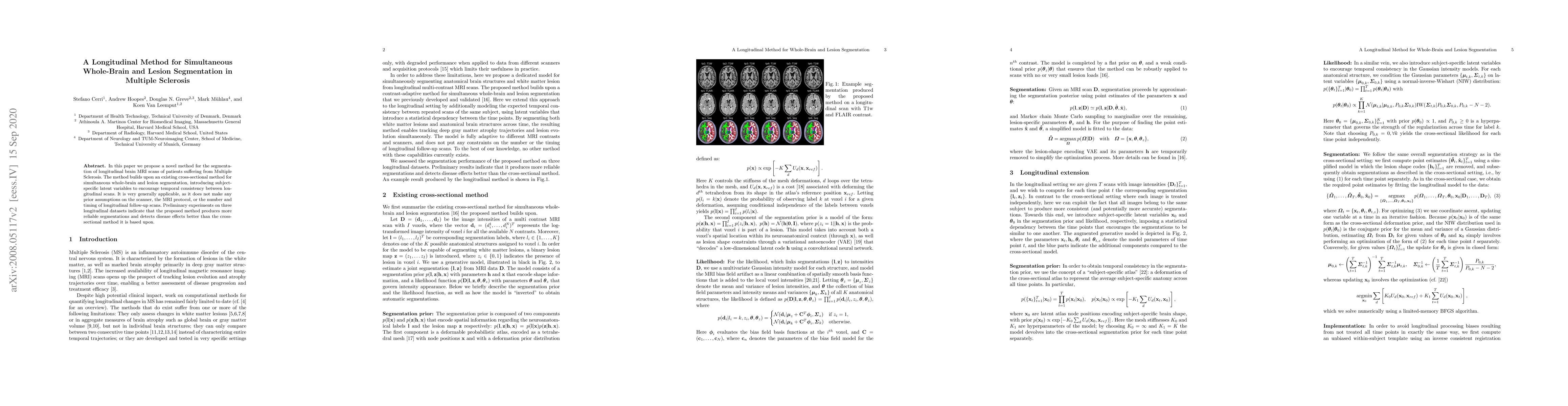
In this paper we propose a novel method for the segmentation of longitudinal brain MRI scans of patients suffering from Multiple Sclerosis. The method builds upon an existing cross-sectional method for simultaneous whole-brain and lesion segmentation, introducing subject-specific latent variables to encourage temporal consistency between longitudinal scans. It is very generally applicable, as it does not make any prior assumptions on the scanner, the MRI protocol, or the number and timing of longitudinal follow-up scans. Preliminary experiments on three longitudinal datasets indicate that the proposed method produces more reliable segmentations and detects disease effects better than the cross-sectional method it is based upon.

Citation & Reference Network Visualization

This graph shows how this paper is connected to other research. The current paper is shown in dark gray, papers that cite it are in green, and papers it references are in blue. You can:

Current Paper
Papers citing this work (Citations)
Papers cited by this work (References)

• Hover over any node to see the paper title

• Click a node to view that paper's details

• Drag nodes to rearrange the graph

• Use the zoom controls (bottom right) to zoom in/out

Citations and References

Title Authors Year Actions
How far MS lesion detection and segmentation are integrated into the clinical workflow? A systematic review
F. Spagnolo, A. Depeursinge, Sabine Schädelin, Aysenur Akbulut, H. Müller, M. Barakovic, L. Melie-García, M. Bach Cuadra, C. Granziera
2023
View Paper
Lesion-Specific Prediction with Discriminator-Based Supervised Guided Attention Module Enabled GANs in Multiple Sclerosis
Jueqi Wang, Derek Berger, Erin Mazerolle, J. Delamer, J. Levman
2022
View Paper
An open-source tool for longitudinal whole-brain and white matter lesion segmentation
Stefano Cerri, D. Greve, Andrew Hoopes, H. Lundell, H. Siebner, Mark Muhlau, K. V. Leemput
2022
View Paper
Normalization of MRI T1w between-scan effects for improved longitudinal volumetric estimates
Donatas Sederevičius, A. Bjørnerud, K. Walhovd, A. Fjell
2022
View Paper
A robust intensity distribution alignment for harmonization of T1w intensity values
Donatas Sederevičius, A. Bjørnerud, K. Walhovd, K. Van Leemput, B. Fischl, A. Fjell
2022
View Paper
BrainFD: Measuring the Intracranial Brain Volume With Fractal Dimension
G. Ashraf, Stylianos Chatzichronis, A. Alexiou, Nikolaos Kyriakopoulos, B. Alghamdi, H. Tayeb, J. Alghamdi, Waseem Khan, Manal Ben Jalal, H. Atta
2021
View Paper
Multiple Sclerosis Lesion Analysis in Brain Magnetic Resonance Images: Techniques and Clinical Applications
Yang Ma, Chaoyi Zhang, M. Cabezas, Yang Song, Zihao Tang, Dongnan Liu, Weidong (Tom) Cai, M. Barnett, Chenyu Wang
2021
View Paper
Deep grey matter injury in multiple sclerosis: A NAIMS consensus statement.
D. Ontaneda, P. Raza, K. Mahajan, D. Arnold, M. Dwyer, S. Gauthier, D. Greve, D. Harrison, R. Henry, David K.B. Li, C. Mainero, W. Moore, S. Narayanan, Jiwon Oh, Raihaan Patel, D. Pelletier, Alexander Rauscher, W. Rooney, N. Sicotte, R. Tam, D. Reich, C. Azevedo
2021
View Paper
A contrast-adaptive method for simultaneous whole-brain and lesion segmentation in multiple sclerosis
Stefano Cerri, O. Puonti, D. Meier, J. Wuerfel, M. Mühlau, H. Siebner, K. V. Leemput
2020
View Paper
Hippocampal volume and affect in response to �uctuating estrogens in menstrual cycle irregularity: A longitudinal single-subject study
Carina Heller, Carina J. Koeppel, Caitlin M. Taylor, Ilona Croy
2023
View Paper
A Systematic Mapping Study of Low-Grade Tumor of Brain Cancer and CSF Fluid Detecting in MRI Images Through Multi-Algorithm Techniques
S. Saeed, Habibullah Bin Haroon, Noor Zaman Jhanjhi, Mehmood Naqvi, Muneer Ahmad
2022
View Paper

Similar Papers

An Open-Source Tool for Longitudinal Whole-Brain and White Matter Lesion Segmentation

Koen Van Leemput, Stefano Cerri, Mark Mühlau, Andrew Hoopes, Douglas N. Greve, Henrik Lundell, Hartwig R. Siebner

Published: 2023-02-13

CoactSeg: Learning from Heterogeneous Data for New Multiple Sclerosis Lesion Segmentation

Jianfei Cai, Hengcan Shi, Zhonghua Wu, Yicheng Wu, Bjoern Picker, Winston Chong

Published: 2023-09-18

Subject-Specific Lesion Generation and Pseudo-Healthy Synthesis for Multiple Sclerosis Brain Images

Wenjia Bai, Berke Doga Basaran, Mengyun Qiao, Paul M. Matthews

Published: 2022-08-04

Comments (0)

This website uses cookies to ensure you get the best experience. Learn more