Summary
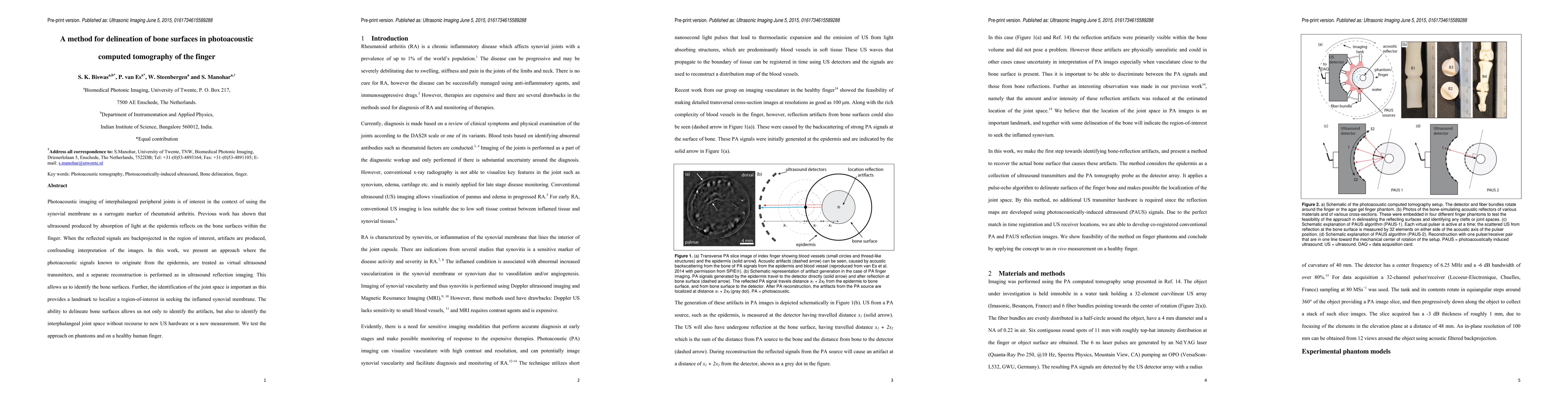
Photoacoustic imaging of interphalangeal peripheral joints is of interest in the context of using the synovial membrane as a surrogate marker of rheumatoid arthritis. Previous work has shown that ultrasound produced by absorption of light at the epidermis reflects on the bone surfaces within the finger. When the reflected signals are backprojected in the region of interest, artifacts are produced, confounding interpretation of the images. In this work, we present an approach where the photoacoustic signals known to originate from the epidermis, are treated as virtual ultrasound transmitters, and a separate reconstruction is performed as in ultrasound reflection imaging. This allows us to identify the bone surfaces. Further, the identification of the joint space is important as this provides a landmark to localize a region-of-interest in seeking the inflamed synovial membrane. The ability to delineate bone surfaces allows us not only to identify the artifacts, but also to identify the interphalangeal joint space without recourse to new US hardware or a new measurement. We test the approach on phantoms and on a healthy human finger.
AI Key Findings
Get AI-generated insights about this paper's methodology, results, and significance.
Paper Details
PDF Preview
Key Terms
Citation Network
Current paper (gray), citations (green), references (blue)
Display is limited for performance on very large graphs.
Similar Papers
Found 4 papersTranscranial photoacoustic computed tomography of human brain function
Yang Zhang, Xin Tong, Li Lin et al.
| Title | Authors | Year | Actions |
|---|
Comments (0)