A Method for Imaging the Ischemic Penumbra with MRI using IVIM
Publication
Metrics
AI Quick Summary
This study demonstrates that intravoxel incoherent motion MRI (IVIM) can effectively quantify local cerebral blood flow and identify the ischemic penumbra in acute ischemic stroke, providing predictive power for infarct volume and growth similar to dynamic susceptibility contrast imaging. IVIM parameters correlate strongly with conventional DSC perfusion metrics and predict final infarct outcomes.
Paper Preview
Abstract
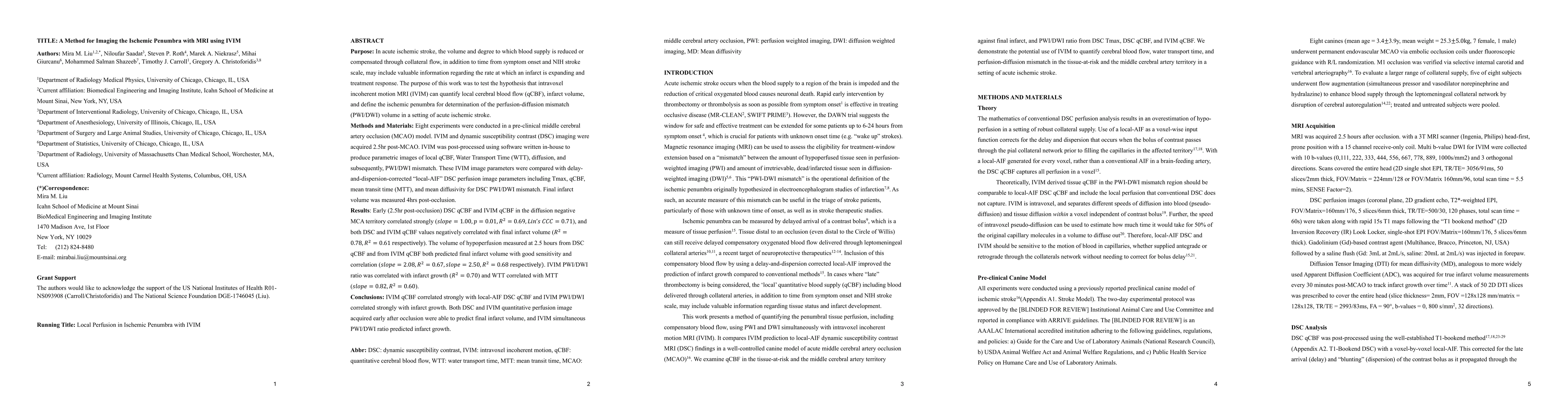
This work examines the hypothesis that intravoxel incoherent motion MRI (IVIM) can quantify local cerebral blood flow (qCBF), infarct volume, and define the ischemic penumbra for determination of the perfusion-diffusion mismatch (PWI/DWI) volume in a setting of acute ischemic stroke. Eight experiments were conducted in a pre-clinical middle cerebral artery occlusion (MCAO) model. IVIM and dynamic susceptibility contrast (DSC) imaging were acquired 2.5hr post-MCAO. IVIM was post-processed using software written in-house to produce parametric images of local qCBF, Water Transport Time (WTT), diffusion, and subsequently, PWI/DWI mismatch. These IVIM image parameters were compared with delay-and-dispersion-corrected local-AIF DSC perfusion image parameters including Tmax, qCBF, mean transit time (MTT), and mean diffusivity for DSC PWI/DWI mismatch. Final infarct volume was measured 4hrs post-occlusion. Early (2.5hr post-occlusion) DSC qCBF and IVIM qCBF in the diffusion negative MCA territory correlated strongly (slope=1.00, p=0.01,R2=0.69,Lins CCC=0.71), and both DSC and IVIM qCBF values negatively correlated with final infarct volume (R2=0.78,R2=0.61 respectively). The volume of hypoperfusion measured at 2.5 hours from DSC qCBF and from IVIM qCBF both predicted final infarct volume with good sensitivity and correlation (slope=2.08, R2=0.67, slope=2.50,R2=0.68 respectively). IVIM PWI/DWI ratio was correlated with infarct growth (R2=0.70) and WTT correlated with MTT (slope=0.82,R2=0.60). IVIM qCBF correlated strongly with local-AIF DSC qCBF and IVIM PWI/DWI correlated strongly with infarct growth. Both DSC and IVIM quantitative perfusion image acquired early after occlusion were able to predict final infarct volume, and IVIM simultaneous PWI/DWI ratio predicted infarct growth.
AI Key Findings
Get AI-generated insights about this paper's methodology, results, significance, and more — seven facets brought into focus.
Impact
Paper Details
Authors
PDF Preview
Citation Network
Current paper (gray), citations (green), references (blue)
Display is limited for performance on very large graphs.
Discussion 0