Summary
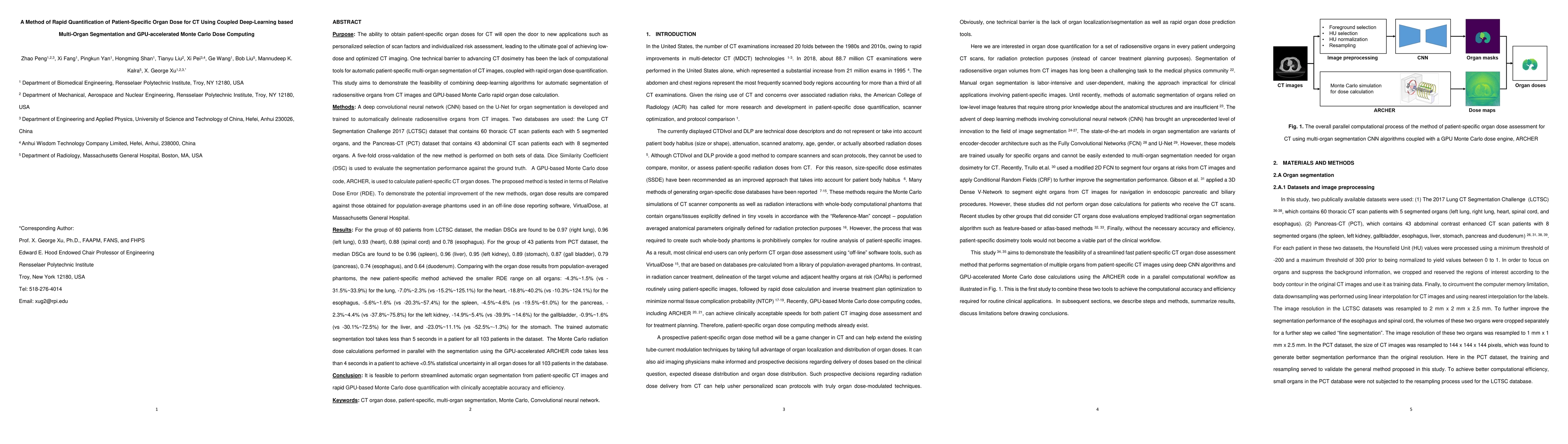
Purpose: This paper describes a new method to apply deep-learning algorithms for automatic segmentation of radiosensitive organs from 3D tomographic CT images before computing organ doses using a GPU-based Monte Carlo code. Methods: A deep convolutional neural network (CNN) for organ segmentation is trained to automatically delineate radiosensitive organs from CT. With a GPU-based Monte Carlo dose engine (ARCHER) to derive CT dose of a phantom made from a subject's CT scan, we are then able to compute the patient-specific CT dose for each of the segmented organs. The developed tool is validated by using Relative Dose Error (RDE) against the organ doses calculated by ARCHER with manual segmentation performed by radiologists. The dose computation results are also compared against organ doses from population-average phantoms to demonstrate the improvement achieved by using the developed tool. In this study, two datasets were used: The Lung CT Segmentation Challenge 2017 (LCTSC) dataset, which contains 60 thoracic CT scan patients each with 5 segmented organs, and the Pancreas-CT (PCT) dataset, which contains 43 abdominal CT scan patients each with 8 segmented organs. Five-fold cross-validation of the new method is performed on both datasets. Results: Comparing with the traditional organ dose evaluation method that based on population-average phantom, our proposed method achieved the smaller RDE range on all organs with -4.3%~1.5% vs -31.5%~33.9% (lung), -7.0%~2.3% vs -15.2%~125.1% (heart), -18.8%~40.2% vs -10.3%~124.1% (esophagus) in the LCTSC dataset and -5.6%~1.6% vs -20.3%~57.4% (spleen), -4.5%~4.6% vs -19.5%~61.0% (pancreas), -2.3%~4.4% vs -37.8%~75.8% (left kidney), -14.9%~5.4% vs -39.9% ~14.6% (gall bladder), -0.9%~1.6% vs -30.1%~72.5% (liver), and -23.0%~11.1% vs -52.5%~-1.3% (stomach) in the PCT dataset.
AI Key Findings
Get AI-generated insights about this paper's methodology, results, and significance.
Paper Details
PDF Preview
Key Terms
Citation Network
Current paper (gray), citations (green), references (blue)
Display is limited for performance on very large graphs.
Similar Papers
Found 4 papersOrgan-Level Radiation Doses from CT Scans for 10,000 Chinese Subjects Undergoing Physical Examinations: The Feasibility of AI-Based Multi-organ CT Image Segmentation and Near Real-time Monte Carlo Dose Computing
Yang Lu, Zhi Chen, Li Tao et al.
Development of a GPU-accelerated Monte Carlo dose calculation module for nuclear medicine, ARCHER-NM: Demonstration for a PET/CT imaging procedure
Yu Lu, Zhi Chen, Bo Cheng et al.
Self is the Best Learner: CT-free Ultra-Low-Dose PET Organ Segmentation via Collaborating Denoising and Segmentation Learning
Zanting Ye, Lijun Lu, Xiaolong Niu et al.
| Title | Authors | Year | Actions |
|---|
Comments (0)