01
MethodologyHow they did it
A deep learning method based on a combination of two UNet-3D branches was proposed for PET/CT lesion segmentation.
This paper proposes a mirror-Unet architecture for segmenting oncologic lesions from [${}^{18}$F]FDG PET/CT scans, combining two UNet-3D branches for CT and PET images to handle diverse lesion characteristics. The method was validated on the AutoPET MICCAI 2023 Challenge dataset, with code available for reproducibility.
A deep learning method based on a combination of two UNet-3D branches was proposed for PET/CT lesion segmentation. More in Methodology →
Mean Dicescore of 0.54 on the preliminary test set — Mean false positive volume of 1.13 ml and mean false negative volume of 0.19 ml More in Key Results →
Automatic lesion detection and segmentation from PET/CT scans is a challenging task, requiring a deep learning method that can handle diverse shapes, sizes, FDG uptake, and location. More in Significance →
TotalSegmentator had better performance in abdominal regions — No normalization of images More in Limitations →
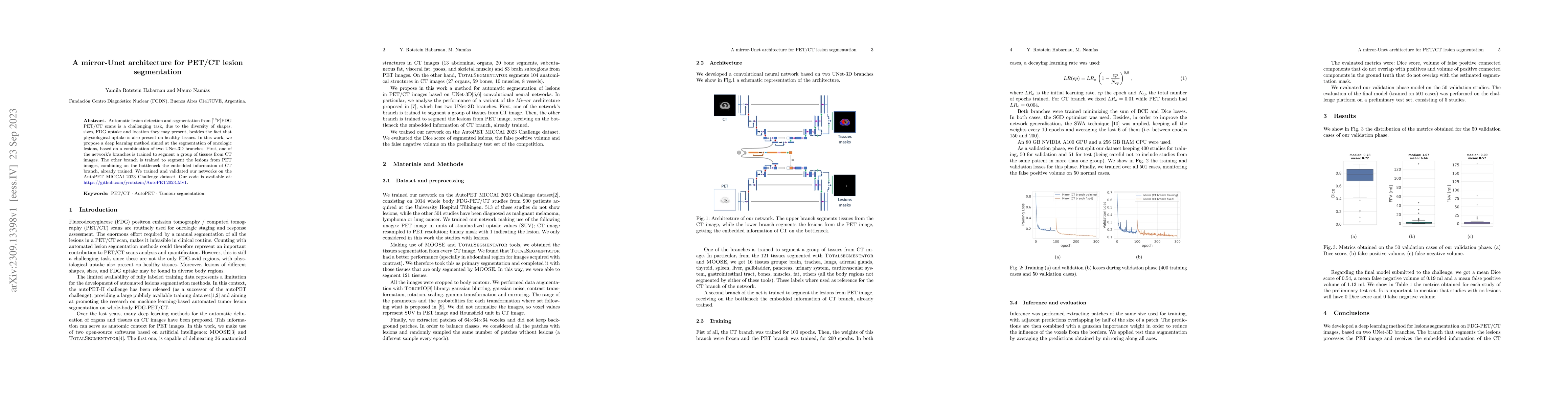
Automatic lesion detection and segmentation from [${}^{18}$F]FDG PET/CT scans is a challenging task, due to the diversity of shapes, sizes, FDG uptake and location they may present, besides the fact that physiological uptake is also present on healthy tissues. In this work, we propose a deep learning method aimed at the segmentation of oncologic lesions, based on a combination of two UNet-3D branches. First, one of the network's branches is trained to segment a group of tissues from CT images. The other branch is trained to segment the lesions from PET images, combining on the bottleneck the embedded information of CT branch, already trained. We trained and validated our networks on the AutoPET MICCAI 2023 Challenge dataset. Our code is available at: https://github.com/yrotstein/AutoPET2023_Mv1.
Seven facets of this paper, analysed and brought into focus by AI.
Automatic lesion detection and segmentation from PET/CT scans is a challenging task, requiring a deep learning method that can handle diverse shapes, sizes, FDG uptake, and location.
A deep learning method based on a combination of two UNet-3D branches was proposed for PET/CT lesion segmentation.
Automatic lesion detection and segmentation from PET/CT scans is a challenging task, requiring a deep learning method that can handle diverse shapes, sizes, FDG uptake, and location.
The proposed mirror-Unet architecture combines two UNet-3D branches to process PET and CT images, respectively.
The use of a bottleneck layer to combine the embedded information of the CT branch with the PET branch is novel and different from existing research
Current paper (gray), citations (green), references (blue)
Display is limited for performance on very large graphs.
Discussion 0