Publication
Metrics
Quick Actions
AI Quick Summary
This paper introduces a model for boundary-driven tissue morphogenesis using the Drosophila hindgut primordium, demonstrating that its shape changes are passive results of surrounding active embryo regions. The study finds that the hindgut's deformations follow an initial translation and a rapid symmetry-breaking, suggesting uniform boundary conditions can generate nonuniform shape changes.
Quick Answers
What is "A model for boundary-driven tissue morphogenesis" about?
This paper introduces a model for boundary-driven tissue morphogenesis using the Drosophila hindgut primordium, demonstrating that its shape changes are passive results of surrounding active embryo regions. The study finds that the hindgut's deformations follow an initial translation and a rapid symmetry-breaking, suggesting uniform boundary conditions can generate nonuniform shape changes.
What methodology did the authors use?
The study utilizes the Drosophila hindgut primordium as a model, characterizing its deformations and constructing a minimal elastic ring model deformed by active midgut invagination and germ band extension on an ellipsoidal surface to capture its triangular shape. More in Methodology →
What are the key results?
Identification of an intermediate triangular shape in 3D deformations of the hindgut. — Discovery of two stages in hindgut deformation: initial translation on the curved embryo surface followed by rapid breaking of shape symmetry. More in Key Results →
Why is this work significant?
This research provides a possible explanation for the various characteristic shapes of blastopore-equivalents in different organisms and offers a framework for understanding the mechanical emergence of global morphologies in complex developmental systems. More in Significance →
What are the main limitations?
The study is limited to the Drosophila hindgut primordium, so findings may not be universally applicable to all organisms. — The model assumes an elastic ring, which might not capture all complexities of biological tissues. More in Limitations →
Abstract
Tissue deformations during morphogenesis can be active, driven by internal processes, or passive, resulting from stresses applied at their boundaries. Here, we introduce the Drosophila hindgut primordium as a model for studying boundary-driven tissue morphogenesis. We characterize its deformations and show that its complex shape changes can be a passive consequence of the deformations of the active regions of the embryo that surround it. First, we find an intermediate characteristic triangular shape in the 3D deformations of the 
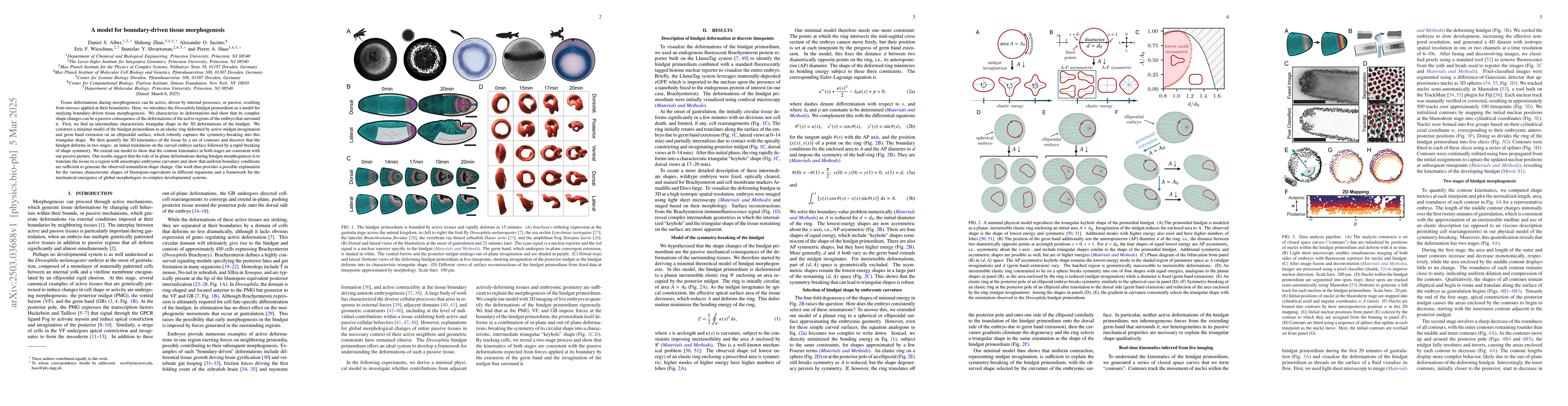
hindgut. We construct a minimal model of the hindgut primordium as an elastic ring deformed by active midgut invagination and germ band extension on an ellipsoidal surface, which robustly captures the symmetry-breaking into this triangular shape. We then quantify the 3D kinematics of the tissue by a set of contours and discover that the hindgut deforms in two stages: an initial translation on the curved embryo surface followed by a rapid breaking of shape symmetry. We extend our model to show that the contour kinematics in both stages are consistent with our passive picture. Our results suggest that the role of in-plane deformations during hindgut morphogenesis is to translate the tissue to a region with anisotropic embryonic curvature and show that uniform boundary conditions are sufficient to generate the observed nonuniform shape change. Our work thus provides a possible explanation for the various characteristic shapes of blastopore-equivalents in different organisms and a framework for the mechanical emergence of global morphologies in complex developmental systems.
AI Key Findings
Generated Jun 10, 2025
Methodology — What approach did the authors take?
The study utilizes the Drosophila hindgut primordium as a model, characterizing its deformations and constructing a minimal elastic ring model deformed by active midgut invagination and germ band extension on an ellipsoidal surface to capture its triangular shape.
Key Results — What are the main findings?
- Identification of an intermediate triangular shape in 3D deformations of the hindgut.
- Discovery of two stages in hindgut deformation: initial translation on the curved embryo surface followed by rapid breaking of shape symmetry.
- Demonstration that in-plane deformations during hindgut morphogenesis translate tissue to a region with anisotropic embryonic curvature under uniform boundary conditions.
Significance — Why does this research matter?
This research provides a possible explanation for the various characteristic shapes of blastopore-equivalents in different organisms and offers a framework for understanding the mechanical emergence of global morphologies in complex developmental systems.
Technical Contribution — What is the technical contribution?
Development of a minimal elastic ring model to explain the triangular shape and deformation stages of the Drosophila hindgut primordium under active and passive forces.
Novelty — What is new about this work?
The research introduces a novel framework for understanding boundary-driven tissue morphogenesis, emphasizing the role of in-plane deformations and anisotropic embryonic curvature in shaping complex developmental structures.
Limitations — What are the limitations of this study?
- The study is limited to the Drosophila hindgut primordium, so findings may not be universally applicable to all organisms.
- The model assumes an elastic ring, which might not capture all complexities of biological tissues.
Future Work — What did the authors propose for future work?
- Investigate the applicability of this model to other organisms and developmental systems.
- Incorporate more complex material properties to better represent biological tissues in the model.
Paper Details
How to Cite This Paper
@article{jacinto2025a,
title = {A model for boundary-driven tissue morphogenesis},
author = {Jacinto, Alexandre O. and Alber, Daniel S. and Wieschaus, Eric F. and others},
year = {2025},
eprint = {2503.03688},
archivePrefix = {arXiv},
primaryClass = {physics.bio-ph},
}
Jacinto, A., Alber, D., Wieschaus, E., Haas, P., Zhao, S., & Shvartsman, S. (2025). A model for boundary-driven tissue morphogenesis. arXiv. https://arxiv.org/abs/2503.03688
Jacinto, Alexandre O., et al. "A model for boundary-driven tissue morphogenesis." arXiv, 2025, arxiv.org/abs/2503.03688.
PDF Preview
Citation Network
Current paper (gray), citations (green), references (blue)
Display is limited for performance on very large graphs.
Similar Papers
Found 4 papersPalaCell2D: A framework for detailed tissue morphogenesis
Bastien Chopard, Raphaël Conradin, Christophe Coreixas et al.
Towards a two-scale model for morphogenesis -- How cellular processes influence tissue deformations
Lea Happel, Axel Voigt
| Title | Authors | Year | Actions |
|---|
Comments (0)