Summary

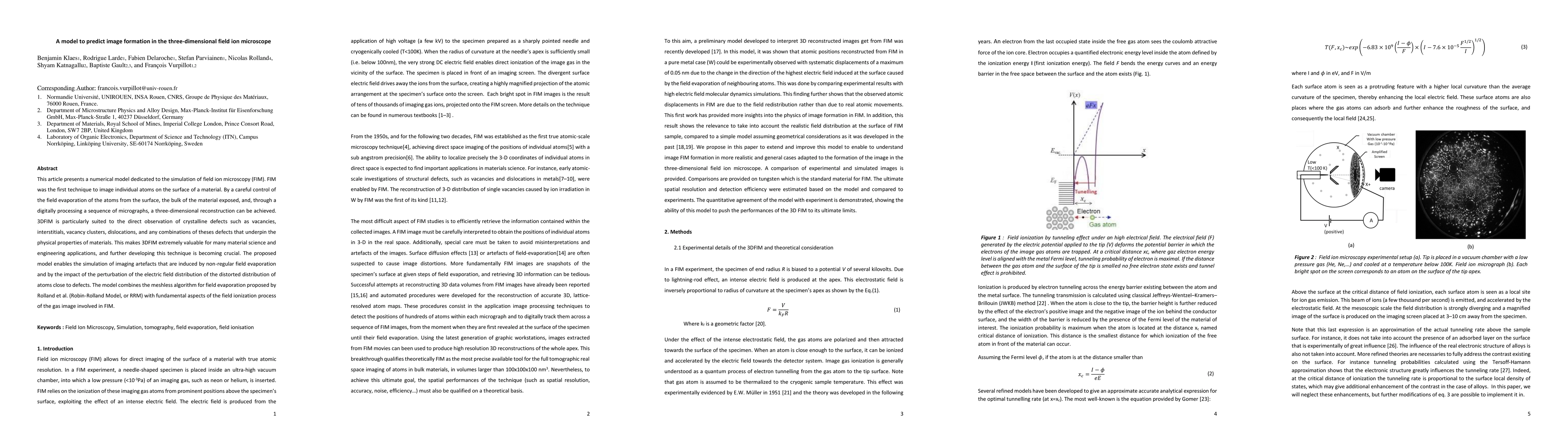
This article presents a numerical model dedicated to the simulation of field ion microscopy (FIM). FIM was the first technique to image individual atoms on the surface of a material. By a careful control of the field evaporation of the atoms from the surface, the bulk of the material exposed, and, through a digitally processing a sequence of micrographs, a three-dimensional reconstruction can be achieved. 3DFIM is particularly suited to the direct observation of crystalline defects such as vacancies, interstitials, vacancy clusters, dislocations, and any combinations of theses defects that underpin the physical properties of materials. This makes 3DFIM extremely valuable for many material science and engineering applications, and further developing this technique is becoming crucial. The proposed model enables the simulation of imaging artefacts that are induced by non-regular field evaporation and by the impact of the perturbation of the electric field distribution of the distorted distribution of atoms close to defects. The model combines the meshless algorithm for field evaporation proposed by Rolland et al. (Robin-Rolland Model, or RRM) with fundamental aspects of the field ionization process of the gas image involved in FIM.

AI Key Findings

Get AI-generated insights about this paper's methodology, results, and significance.

Paper Details

PDF Preview

Download PDF

Key Terms

simulation (0.152)
model (0.116)
proposed (0.111)
particularly (0.103)
enables (0.100)
impact (0.095)
numerical (0.078)
non (0.061)

Citation Network

Current paper (gray), citations (green), references (blue)

Current Paper
Citations
References
Click to view
Title Authors Year Actions
On the Iron Content of Mn-Ni-Si-rich Clusters That Form in Reactor Pressure Vessel Steels During Exposure to Neutron Irradiation
Benjamin M. Jenkins, Aidar K. Zakirov, François Vurpillot, Auriane Etienne, C. Pareige, P. Pareige, Bertrand Radiguet
2024
View Paper
Surface Dynamics of Field Evaporation in Silicon Carbide
S. Ndiaye, C. Bacchi, B. Klaes, M. Canino, F. Vurpillot, L. Rigutti
2023
View Paper
Accurate computation of chemical contrast in field ion microscopy
Shalini Bhatt, Shyam Katnagallu, J. Neugebauer, C. Freysoldt
2022
View Paper
Identification of Crystalline Orientation of Tungsten Tips by Machine Learning Analysis of Field Ion Micrographs
M. Yamada, Tadasuke Okazawa, S. Nagai, K. Hata
2022
View Paper
Atom Probe Tomography Analysis of Mica
Chiara Cappelli, A. Pérez-Huerta, S. B. Alam, T. Prozorov
2021
View Paper
Analytical Three-Dimensional Field Ion Microscopy of an Amorphous Glass FeBSi
B. Klaes, Jeoffrey Renaux, R. Lardé, F. Delaroche, F. Morgado, L. Stephenson, B. Gault, F. Vurpillot
2021
View Paper
Atom probe tomography
B. Gault, A. Chiaramonti, O. Cojocaru-Mirédin, P. Stender, R. Dubosq, C. Freysoldt, S. Makineni, Tong Li, M. Moody, J. Cairney
2021
View Paper
Three-Dimensional Atomically Resolved Analytical Imaging with a Field Ion Microscope
Shyam Katnagallu, F. Morgado, I. Mouton, B. Gault, L. Stephenson
2021
View Paper
Development of Wide Field of View Three-Dimensional Field Ion Microscopy and High-Fidelity Reconstruction Algorithms to the Study of Defects in Nuclear Materials
B. Klaes, R. Lardé, F. Delaroche, C. Hatzoglou, Stefan Parvianien, J. Houard, G. Da Costa, A. Normand, Martin Brault, B. Radiguet, F. Vurpillot
2021
View Paper
Reflections on the Spatial Performance of Atom Probe Tomography in the Analysis of Atomic Neighborhoods
B. Gault, B. Klaes, F. Morgado, C. Freysoldt, Yue Li, F. De Geuser, L. Stephenson, F. Vurpillot
2021
View Paper
Revealing atomic-scale vacancy-solute interaction in nickel
F. Morgado, Shyam Katnagallu, C. Freysoldt, B. Klaes, F. Vurpillot, J. Neugebauer, D. Raabe, S. Neumeier, B. Gault, L. Stephenson
2021
View Paper
Understanding field evaporation sequences by in-situ correlative microscopy and simulation
Mohammed Ilhami, Williams Lefebvre, François Vurpillot
2024
View Paper

Comments (0)

This website uses cookies to ensure you get the best experience. Learn more