This paper introduces a new fiber-based cathodoluminescence detector with 3D printed micro-optics for scanning electron microscopes, demonstrating high spatial resolution and spectral excitation bandwidth. The study validates the system's ability to analyze the spatial and spectral characteristics of emitted light from electron-matter interactions in nanostructures.
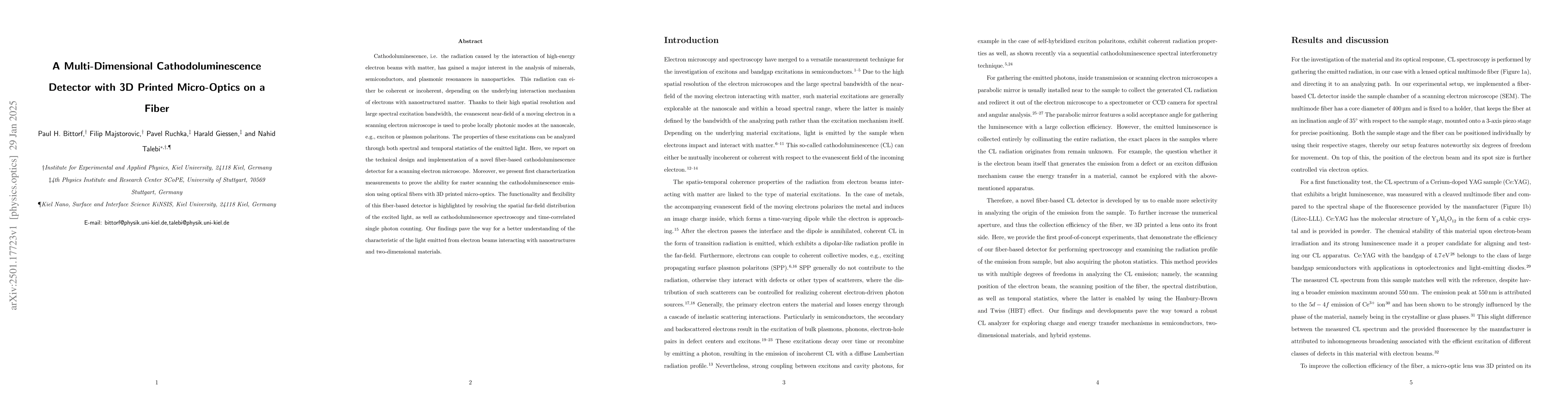
Cathodoluminescence, i.e. the radiation caused by the interaction of high-energy electron beams with matter, has gained a major interest in the analysis of minerals, semiconductors, and plasmonic resonances in nanoparticles. This radiation can either be coherent or incoherent, depending on the underlying interaction mechanism of electrons with nanostructured matter. Thanks to their high spatial resolution and large spectral excitation bandwidth, the evanescent near-field of a moving electron in a scanning electron microscope is used to probe locally photonic modes at the nanoscale, e.g., exciton or plasmon polaritons. The properties of these excitations can be analyzed through both spectral and temporal statistics of the emitted light. Here, we report on the technical design and implementation of a novel fiber-based cathodoluminescence detector for a scanning electron microscope. Moreover, we present first characterization measurements to prove the ability for raster scanning the cathodoluminescence emission using optical fibers with 3D printed micro-optics. The functionality and flexibility of this fiber-based detector is highlighted by resolving the spatial far-field distribution of the excited light, as well as cathodoluminescence spectroscopy and time-correlated single photon counting. Our findings pave the way for a better understanding of the characteristic of the light emitted from electron beams interacting with nanostructures and two-dimensional materials.
Generated Jun 13, 2025
The research utilized a fiber-based cathodoluminescence (CL) detector integrated with 3D printed micro-optics on a fiber for a scanning electron microscope (SEM). This setup allowed for raster scanning of CL emission, resolving spatial far-field distribution, and performing CL spectroscopy and time-correlated single-photon counting.
This research contributes to a better understanding of the characteristics of light emitted from electron beams interacting with nanostructures and two-dimensional materials, which is crucial for advancing nanoscale photonics, electron microscopy, and materials science.
The novel fiber-based CL detector with 3D printed micro-optics on a fiber enhances the collection efficiency and flexibility of CL measurements in SEM, enabling advanced spectroscopy and time-correlated single-photon counting.
This work introduces a versatile fiber-based CL detector with integrated 3D printed micro-optics, which improves light collection efficiency and enables advanced CL measurements, distinguishing it from previous CL detection methods.
Esben Ravn Andresen, Siddharth Sivankutty, Hervé Rigneault et al.
Yanis Taege, Tim Samuel Winter, Sophia Laura Schulz et al.
Juejun Hu, Tian Gu, Luigi Ranno et al.
No citations found for this paper.
Comments (0)