A Multi-Scale Conditional Deep Model for Tumor Cell Ratio Counting
Publication
Metrics
Paper Preview
Abstract
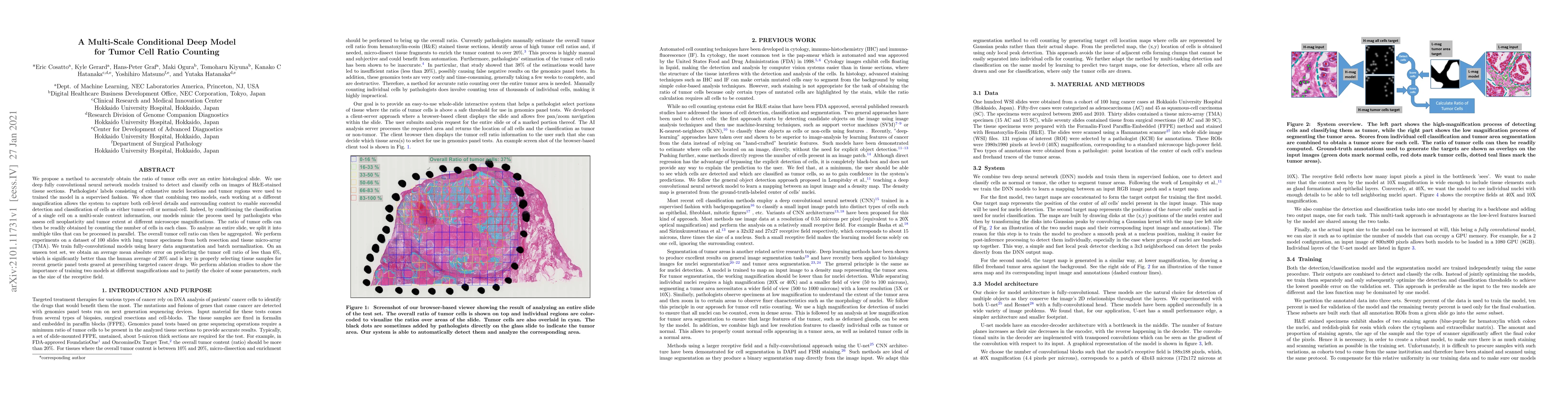
We propose a method to accurately obtain the ratio of tumor cells over an entire histological slide. We use deep fully convolutional neural network models trained to detect and classify cells on images of H&E-stained tissue sections. Pathologists' labels consisting of exhaustive nuclei locations and tumor regions were used to trained the model in a supervised fashion. We show that combining two models, each working at a different magnification allows the system to capture both cell-level details and surrounding context to enable successful detection and classification of cells as either tumor-cell or normal-cell. Indeed, by conditioning the classification of a single cell on a multi-scale context information, our models mimic the process used by pathologists who assess cell neoplasticity and tumor extent at different microscope magnifications. The ratio of tumor cells can then be readily obtained by counting the number of cells in each class. To analyze an entire slide, we split it into multiple tiles that can be processed in parallel. The overall tumor cell ratio can then be aggregated. We perform experiments on a dataset of 100 slides with lung tumor specimens from both resection and tissue micro-array (TMA). We train fully-convolutional models using heavy data augmentation and batch normalization. On an unseen test set, we obtain an average mean absolute error on predicting the tumor cell ratio of less than 6%, which is significantly better than the human average of 20% and is key in properly selecting tissue samples for recent genetic panel tests geared at prescribing targeted cancer drugs. We perform ablation studies to show the importance of training two models at different magnifications and to justify the choice of some parameters, such as the size of the receptive field.
AI Key Findings
Get AI-generated insights about this paper's methodology, results, significance, and more — seven facets brought into focus.
Impact
Paper Details
Authors
PDF Preview
Key Terms
Citation Network
Current paper (gray), citations (green), references (blue)
Display is limited for performance on very large graphs.
Discussion 0