01
MethodologyHow they did it
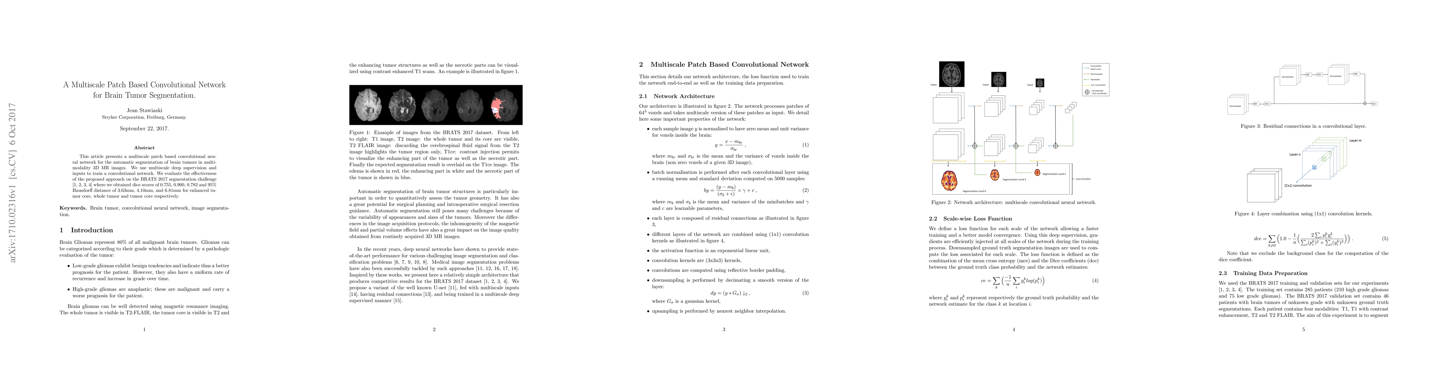
A multiscale patch based convolutional neural network is proposed for automatic brain tumor segmentation in multi-modality 3D MR images.
Discussion 0